Immunoassays
Promega immunoassay reagents and antibody-based detection systems include novel Lumit® Immunoassays, as well as a range of primary and secondary antibodies and detection reagents used in traditional ELISA or Western blotting techniques.
Lumit® Immunoassays provide a simple and fast alternative to conventional immunoassay methods including sandwich ELISAs and Western blots. The bioluminescence-based method can be completed in as little as 30 minutes. We provide several types of Lumit® Immunoassay for research use, including specific kits to detect protein phosphorylation, cytokines, metabolites or neonatal Fc receptor binding. Lumit® detection reagents and labeling kits to build your own Lumit® assays are also available.
Immunoassay Product Groups
Lumit® Immunoassays for Research
A simple, rapid and sensitive alternative to ELISAs and other conventional immunoassays.
Antibody Labeling
Kits for labeling antibodies with bioluminescent tags or fluorescent dyes.
Primary and Secondary Antibodies
Antibodies, substrates and accessory products for immunoblotting, immunocytochemistry and immunohistochemistry.
Top Immunoassay Products for Your Lab
Lumit® Immunoassay Labeling Kit and Detection Reagents
Antibody labeling kit and detection reagents to help develop Lumit® Immunoassays.
VB2500, VB2010, VB2020, VB2030, VB4050, VB4060
Lumit® Immunoassay Cellular Systems
A no-wash bioluminescent immunoassay to measure levels of target analytes in cell lysates.
W1201, W1202, W1203, W1331, W1332, W1333, W1220
Lumit® SARS CoV-2 Spike RBD: hACE2 Immunoassay
A homogeneous bioluminescent assay that measures interaction between SARS-CoV-2 spike protein receptor binding domain (RBD) and human angiotensin-converting enzyme 2 (ACE2).

What are Immunoassays?
Immunoassays are convenient and widely used detection methods based on the ability of antibodies to bind and detect specific antigens. ELISA (Enzyme Linked Immunosorbent Assays) and Western blot techniques are typical examples of commonly used immunoassays.
ELISAs are performed in multiwell plates in several formats. In a sandwich ELISA, an antibody is bound to the plate surface and used to capture a target antigen from a sample such as plasma or serum. After thorough washing, a second antibody is added, which binds to the antigen, creating an antibody-antigen-antibody “sandwich”. This second antibody may be labeled with an enzyme substrate that enables detection, or it may be detected with a labeled species-specific secondary antibody (e.g., anti-human IgG). In other ELISA formats, the target antigen may be bound directly to the plate and used to detect antibodies present in a sample. These bound antibodies are then detected using a secondary labeled antibody.
In an enzyme immunoassay, the enzyme linked to the detecting antibody generates a color signal proportional to the amount of target antigen present in the original sample. Depending on the immunoassay format, the degree of color can be detected and measured using a spectrophotometric or fluorescence plate reader.
In Western blotting, individual proteins present in a sample are separated on a gel and transferred onto a nitrocellulose membrane, which is then interrogated with labeled antibodies directed against specific antigens. After washing, the bound antibodies are visualized by development of the signal (color, fluorescence or chemiluminescence) generated by the labeled primary antibody or a secondary antibody.
ELISA and Western blot techniques offer advantages of specificity, sensitivity and relative ease of use. However, they are time-consuming to perform, involving numerous wash and incubation steps. The number of steps involved can also lead to significant assay-to-assay variability.
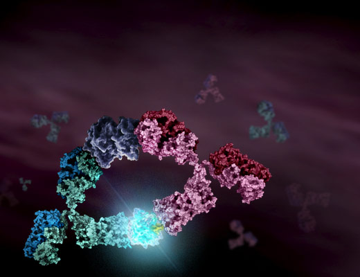
Lumit® Immunoassays differ from Western blots and ELISAs by providing a no-wash, bioluminescence-based method for detecting various target molecules in a rapid and sensitive format. Lumit® Immunoassays are based on NanoLuc® Binary Technology (NanoBiT®). In Lumit® Immunoassays, antibodies or antigens are chemically labeled with the small and large subunits of NanoBiT® luciferase, known as SmBiT and LgBiT, respectively. In the presence of the target molecule, the two subunits are brought into close proximity, allowing SmBiT and LgBiT to form an active enzyme and generate a bright luminescence signal, which is detected using a microplate reader.