Bioluminescence Technology
Bioluminescence is widely used in research because it provides a sensitive, low-background signal for monitoring biological processes. From reporter assays and cell viability to protein detection, immunoassays, and live-cell imaging, bioluminescent tools offer unmatched versatility across the full range of cellular and biochemical questions.
See What Others Can't. Measure What Others Miss.
Bioluminescence-based tools deliver sensitivity, specificity and signal clarity that fluorescence-based methods simply can't match. And for more than three decades, we have been at the center of developing tools that define how researchers measure, detect and visualize biology.
Interested in luciferase reporter assays, including dual formats, but not sure which luciferase reporter system to choose?
Better Tools Mean Better Questions
Not every assay lets you see what you're actually looking for. When your assay has the sensitivity, range and flexibility you need, you stop working around your tools and start getting real answers.
Sensitivity
A primary advantage of bioluminescent assays is that they are generally very sensitive. Few eukaryotic cells produce their own light, so emission from a bioluminescent reaction, or signal strength, is much higher than any background signal. Bioluminescent assays, therefore, have very high signal-to-background ratios—bioluminescence can be routinely measured to levels as low as 10–20 moles. For a typical biological sample, that is equivalent to detecting only a few molecules per cell. This level of sensitivity is often compatible with endogenous expression levels, allowing for bioluminescent tools to accurately reflect native cellular biology.
Dynamic Range
The bright signal of bioluminescence assays allows for measuring a large dynamic range of bioluminescent emissions. Most bioluminescent assays give linear responses over six to eight orders of magnitude of substrate concentration. Fortunately, luminometers, the instruments of choice for quantifying bioluminescence, are capable of operating over these very large ranges.
High-Throughput Assays
The large linear dynamic range and corresponding sensitivity of bioluminescence allows for assays to be miniaturized and performed in medium to high-throughput plate formats, even 1536-well plates. Native bioluminescent reactions have been optimized by our scientists to enable quantitative light-based measurements that use “add-mix-measure” methods that simplify high-throughput assay formats.
Flexible Assay Design
The native firefly bioluminescent reaction gives a brief flash of light with a half-life of about 2 minutes. Our scientists have developed systems that yield longer-lived bioluminescent signals. In our flash-type assays, the signal half-life may only be about 10 minutes, but the signal is relatively bright. Our glow-type assays, in contrast, are less intense, but have signal half-lives often lasting several hours. These reagents can also be added directly to cells in culture, allowing for “add-mix-measure” assays that eliminate sample preprocessing and that are compatible with low- to high-throughput assay workflows.
Signal can be Coupled to a Range of Processes
Another benefit offered by bioluminescence assays is that light intensity generated by bioluminescent reactions is dependent on the concentrations of the component reactants. By coupling biological processes to these components, bioluminescent signals can be associated with a variety of molecular processes, including cell death, gene expression, protein-protein interactions and kinase activity.

Wondering how bioluminescent assays could help answer questions in your research?
The assay you choose determines the reproducibility and defensibility of your data. Talk with one of our scientists to learn what your options are. We have more than 30 years’ experience with bioluminescence and bioluminescent assays.
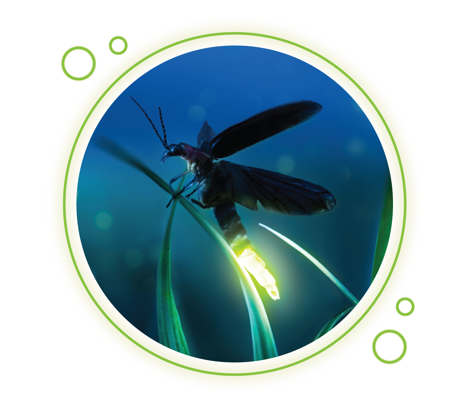
What is Bioluminescence?
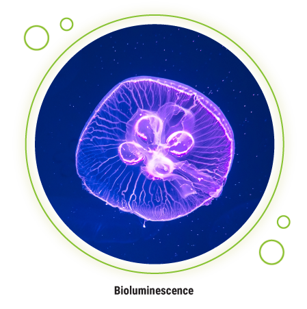
Bioluminescence is a type of luminescence found in biological organisms where light is emitted due to a naturally catalyzed chemical reaction. These light-yielding reactions show up in organisms as diverse as fireflies, jellyfish, bacteria and more.
There are other types of luminescence as well, such as radioluminescence, which relies on radioactivity (e.g., scintillants) to produce light; and electroluminescence, which relies on electricity.
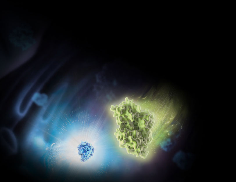
How Does Bioluminescence Work?
In a bioluminescent reaction, light is generated from chemical energy. A good example is the reaction catalyzed by the enzyme luciferase, which is the same reaction that makes fireflies glow. This reaction involves luciferin, oxygen (O2) and adenosine triphosphate (ATP) as the reagents. Luciferase and Mg2+ are catalysts for the reaction. Luciferase catalyzes the oxidation of luciferin to oxyluciferin. During the reaction, luciferin undergoes excitation to a more energetic chemical state. Then, the compound undergoes vibrational relaxation to a lower energy state. A final relaxation to ground state oxyluciferin releases energy in the form of light, a process called emission.


This luciferase-luciferin reaction is a canonical example of a native bioluminescent system. In many systems, luciferase is the enzyme and luciferin is the substrate; their chemical reaction produces light emission that can be quantified as luciferase activity. However, the native luciferase gene is not always optimal for use as a reporter gene in genetic reporter assays or other biological assays. In the 1990s, our scientists used genetic engineering to steadily optimize the native firefly luciferase gene for reporter applications. The final design offered a system where luciferase protein expresses uniformly and optimally in mammalian host cells, minimizes off-target responses and responds rapidly to transcriptional dynamics driven by a promoter.
Other substrate and enzyme pairings can also generate bioluminescence, and different luciferase proteins are used across applications. The color, intensity and duration of the emitted light will vary depending on the thermodynamics and kinetics of the specific reaction. For example, our scientists used directed .
The luminescent signal that results from a luminescent reaction can be coupled to any of the other reagents in the reaction. As an example, in a genetic reporter assay, the gene coding for luciferase is put under the control of the regulatory sequences from a gene of interest. When a reagent containing luciferin and ATP are added to the cell, the amount of light produced will be proportional to the amount of luciferase expressed. Higher gene expression results in greater luciferase expression and luminescent signal and vice versa. Other assays can measure the quantity or changes in ATP concentration or enzymatic release of luciferin or other luminescent substrates.
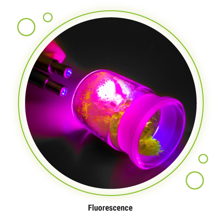
What’s the Difference Between Bioluminescence and Fluorescence?
Bioluminescence and fluorescence are both natural phenomena and are both regularly used in the lab as tools for characterizing biological systems and cellular activity. The main difference between bioluminescence and fluorescence is the excitation source. Whereas chemical energy drives bioluminescence, photons drive fluorescence.
Fluorescent emissions can be very bright compared to luminescent emissions because the photons that drive excitation to the excited states can be introduced to a system at high rates. However, in biological assays, this influx of photons can also lead to higher backgrounds due to two factors: the need for the light detector to discriminate between excitation and emission photons and interference from other fluorescent groups in the sample.
For some techniques, like cellular microscopy and flow cytometry, instrument optics can restrict light so high backgrounds are not a problem. In these cases, the brightness of the system is the most important factor, and fluorescence is most commonly used.
For other cases where optical detection instruments are simpler and where there are more samples to analyze, lower background is needed. In those cases, luminescence can be a more valuable tool for biological assays and for studying cell activity with fewer optical artifacts. Because the readout is based on an enzyme-driven reaction rather than excitation light, luminescence assays are well suited for monitoring promoter activity and transcription with low background in live cell or lysate formats.
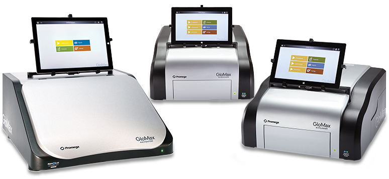
How Do You Measure Luminescence?
A luminometer is the most used instrument for measuring luminescence. They’re often simpler than instruments used to measure absorbance or fluorescence since they do not need a light source or filter, only a light detector. Luminometers usually don’t need to have a specific detection wavelength selected for an assay—they'll just collect light across the entire visible spectrum. This contributes to the strong signal-to-background you can achieve with luminescent assays.
There are two additional pieces of equipment you’ll need to carry out a luminescent assay.
If you’re using a microplate reader to analyze your assay, then you’ll need opaque white microplates, which reflect light and will maximize your assay’s signal. The bottoms of the wells can also be opaque or clear.
If you’re using a luminescent assay that produces a “flash-type” signal instead of a “glow-type” signal, then you’ll need one more piece of equipment—an injector. An injector will add substrate required to initiate a luminescent reaction and will coordinate the injection with signal collection from the luminometer.
Explore Luminometer Options
*For Research Use Only. Not for Use in Diagnostic Procedures.
Ask More Questions, Get More Answers
We've developed a variety of luminescent assays that can answer your research questions. Each innovation made something measurable that wasn't before.
Luciferase Reporter Assay Compairson Guide
Assays for Biochemical Targets
ATP Assays for Measuring Cell Viability
Antibody-Free Protein Detection with HiBiT Protein Tagging Technology
Bioluminescent Immunodetection with Lumit® Immunoassays
NanoLuc® Luciferase for Diverse and Sensitive Bioluminescent Assays
Want to see the potential of bioluminescence in action? Watch these videos to see how scientists transformed their research using bioluminescent assays.
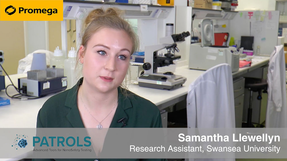
Developing Physiologically Relevant 3D Liver Models for HT Screening
Dr. Samantha Llewellyn at Swansea University uses the CellTiter-Glo® 3D assay to study her 3D cellular liver models. Learn more about bioluminescent assays optimized for use with 3D cell culture models with our Introduction to 3D Cell Culture guide.

From Live Cells to Lysates: Adapting NanoBiT to a Biochemical Assay Format
Dr. Mohammad Ismail at the Francis Crick Institute uses a NanoBiT® Protein:Protein Interaction assay in a novel way to detect small molecule inhibitors for cancer drug discovery targets. Learn more about how our NanoBiT® technology was adapted to a biochemicalassay.
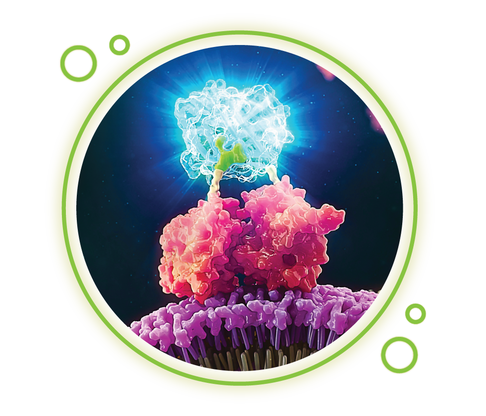
See the Biology Behind the Signal
Traditional luminescence plate readers tell you a process occurred. They can't tell you where in the cell, or how it changed over time. Live-cell bioluminescence imaging closes that gap by turning invisible dynamics into visible, measurable events. Follow protein–protein interactions, target engagement, and other cellular processes in real time, in living cells, without sacrificing the sensitivity that makes bioluminescence the right tool for the question.
*For Research Use Only. Not for Use in Diagnostic Procedures.