Apoptosis
Learn the molecular mechanisms of apoptosis and how to detect, confirm, and measure it using single-sample multiplexed assays.
Promega Corporation
Publication date: 2026
Key Takeaways
- Apoptosis is programmed cell death mediated by caspase proteases, activated via extrinsic (death receptor) or intrinsic (mitochondrial) pathways.
- Caspase activity is a primary marker for detecting apoptosis; timing of apoptotic markers occurs in waves, with initiator caspases (8, 9) activating effector caspases (3, 7).
- Detection methods include caspase activity assays, DNA fragmentation (TUNEL), mitochondrial markers, and annexin V binding.
- More than one method is usually needed to confirm apoptosis, especially useful for distinguishing apoptosis from other regulated modes of cell death.
- Multiplexing assays on a single sample adds internal controls and eliminates repeated work.
What Is Apoptosis?
Apoptosis is programmed cell death— a tightly regulated, genetically encoded process distinct from necrosis. It proceeds through characteristic morphological and biochemical changes, occurs without provoking inflammation, and affects individual cells rather than groups. Understanding apoptosis is central to drug discovery and disease research.
Cell Death as Cell Fate: Historical Context
Although apoptosis is often described as a "hot topic" or a "new and exploding" area of biological research, the concept of cell death as a normal cell fate was articulated only three years after Schleiden and Schwann introduced the Cell Theory when, in 1874, Vogt described natural cell death as an integral part of toad development (Cotter and Curtin, 2003). In 1885 Flemming provided the first morphological description of a natural cell death process, which we now label "apoptosis", a term coined by Kerr and colleagues to describe the unique morphology associated with a cell death that differs from necrosis (Kerr et al. 1972).
Studies of cell lineage in the nematode Caenorhabditis elegans showed that apoptosis was a normal feature of the nematode's invariant developmental program. Of the 1,090 somatic cells of the C. elegans adult hermaphrodite, 131 die during normal development (Hengartner, 1997; Mustafa et al. 2024), and the lineage and timing of apoptosis for each of these cells were constant—demonstrating that apoptosis is genetically programmed. At the biochemical level, Wyllie showed that DNA degradation by a specific endonuclease during apoptosis resulted in a DNA ladder composed of mono- and oligonucleosomal-sized fragments (Wyllie, 1980).
Morphology and Overview of Apoptosis
Morphologically, apoptosis is first characterized by a change in the refractive index of the cell (Hengartner, 1997; Mustafa et al. 2024) followed by cytoplasmic shrinkage and nuclear condensation. The cell membrane begins to show blebs or spikes, and eventually these separate from the dying cell to form "apoptotic bodies" (Figure 1). Apoptotic cells cease to maintain phospholipid asymmetry, and phosphatidylserine (PS) appears on the outer leaflet (Williamson, 2000). The mitochondrial outer membrane (MOM) loses its electrochemical gradient, and substances such as cytochrome c leak into the cytoplasm. Adjacent cells or macrophages phagocytose the apoptotic bodies. The apoptotic cell does not provoke an inflammatory response, and only individual cells are affected in vivo.
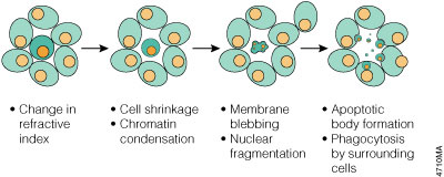
Figure 1. Morphology changes during apoptosis. The cell membrane begins to show blebs or spikes, depending on cell type. Eventually these separate from the dying cell and form "apoptotic bodies" that are phagocytosed by neighboring cells.
Apoptosis is one of several regulated death modes. Take necrosis for example, which is first marked by loss of cell membrane integrity. The cytoplasm and mitochondria swell and lyse, with no vesicles or apoptotic bodies forming and groups of adjacent cells often being affected. Necrotic cell remnants are phagocytosed by macrophages and inflammatory responses are provoked in vivo.
Although apoptosis and necrosis represent distinct mechanism-based modes of cell death, they can overlap morphologically during late stages. Caspase activation is the hallmark of apoptotic commitment, though it has been shown that cells can undergo caspase independent cell death (CICD) when mitochondrial outer membrane permeabilization occurs, or via reactive oxygen species (ROS) mechanisms (Chen, 2022; Gupta, 2025). In vitro, apoptotic cells that are not cleared eventually lose membrane integrity and undergo "secondary necrosis" (Riss and Moravec, 2004). Early frameworks described apoptosis-like and necrosis-like programmed cell death; current knowledge requires multiplexed analysis of several markers to appropriately distinguish modes of cell death (Leist & Jäättelä, 2001). Because apoptosis progresses through distinct, transient stages, measuring multiple markers at sequential time points is recommended.
Apoptosis vs. Necrosis at a Glance
| Feature | Apoptosis | Necrosis |
|---|---|---|
| Membrane integrity | Maintained, then blebbing | Lost early |
| Inflammation | None in vivo | Provoked in vivo |
| Cells affected | Individual cells | Groups of adjacent cells |
| Organelles | Shrinkage, nuclear condensation | Swelling, lysis |
What Are Caspases and What Role Do They Play in Apoptosis?
Caspases are a conserved family of cysteine proteases that drive apoptosis. They exist as inactive zymogens activated by proteolytic cleavage. Initiator caspases (caspase-8 and -9) respond to apoptotic signals and activate effector caspases (caspase-3 and -7), which cleave downstream targets to execute cell death. There are also caspase independent death pathways, as highlighted above.
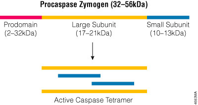
Figure 2. The active caspase enzyme. Caspase zymogens are cleaved between the large and small subunits, and the prodomains are removed. The active site is formed by a heterodimer that contains one large and one small subunit. Two heterodimers associate to form the fully active tetramer.
What Is the Role of Bcl-2 Family Proteins in Apoptosis?
Bcl-2 family proteins regulate the mitochondrial (intrinsic) pathway. Anti-apoptotic members (Bcl-2, Bcl-XL) protect cells by sequestering pro-apoptotic proteins, while pro-apoptotic members (Bax, Bad, Bid) permeabilize the mitochondrial outer membrane, releasing cytochrome c. Their balance determines whether a cell commits to apoptosis (Kaloni et al. 2023).
How Is Apoptosis Activated?
The extrinsic pathway is triggered by cell surface "death receptors" (Fas, TNFR1, TRAIL-R1/R2, p75-NGFR) that carry death domains (DD) and death effector domains (DED). It typically activates initiator caspase-8, which either activates caspase-3 or cleaves the Bcl-2 family member Bid, leading to apoptosome formation and caspase-9 activation (Hengartner, 2000; Wajant et al. 2003). The intrinsic (mitochondrial) pathway is activated by stimuli independent of death receptors—DNA damage, topoisomerase inhibition, or trophic factor withdrawal—and involves Bcl-2 family members regulating cytochrome c release (Rich et al. 1999; Parone et al. 2003). A third route to the intrinsic pathway begins at the endoplasmic reticulum. Sustained ER stress and disrupted protein homeostasis can push a cell toward apoptosis, and in human cells caspase-4 has been proposed as a participant in this response. Caspase-12, first identified in rodents as an ER-stress mediator, is now understood differently in humans: the CASP12 gene is nonfunctional in most people, and where the full-length protein is expressed, it acts in inflammatory signaling rather than as an executioner of ER-stress apoptosis (Wang et al. 2006; Science Direct, 2002).
Fas: An Example of the Death Receptor Pathway
Fas receptor aggregation upon binding multivalent Fas Ligand (FasL) assembles the death inducing signaling complex (DISC; Figure 3) at the cytoplasmic tail of the receptors. The DISC recruits the adaptor protein FADD and procaspase-8; the resulting high local concentration of procaspase-8 permits activation (induced proximity activation; Hengartner, 2000), though other work indicates dimerization is required (Boatright et al. 2003). Active caspase-8 then activates effector caspase-3. In some cells caspase-8 also cleaves Bid, inducing Bax-mediated cytochrome c release from the mitochondria and amplifying the death signal.
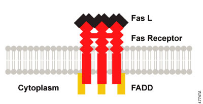
Figure 3. DISC Formation. Aggregation of activated death receptors brings the cytoplasmic domains into close proximity and induces a conformational change that allows the assembly of the death inducing signaling complex (DISC) at the cytoplasmic tail of the receptors.
The Mitochondrial Pathway (Intrinsic)
The mitochondrial pathway involves Bcl-2 family proteins and can be activated by the death receptor pathway or by DNA damage, topoisomerase inhibition, or trophic factor withdrawal (Parone et al. 2003). Pro-apoptotic Group II/III members (Bax, Bad, Bid) shuttle to the mitochondria and interact with anti-apoptotic members (Bcl-2, Bcl-XL) to determine whether apoptosis proceeds. If pro-apoptotic proteins prevail, cytochrome c is released and interacts with Apaf-1 (homolog of C. elegans CED-4; Zou et al. 1997), dATP and procaspase-9 to form the apoptosome, activating caspase-9 and then caspase-3.
Clinical Applications of Apoptosis Research
Many diseases—cancers, autoimmune diseases and neurodegenerative diseases (Alzheimer's, Huntington's, ALS)—reflect either a failure of apoptosis to eliminate harmful cells or the inappropriate activation of apoptosis (Murphy et al. 2003; Rajesh & Kanneganti, 2022). This has made the apoptotic pathway a promising and sustained drug target. The clearest success targeting this pathway is in oncology. One example of a BH3 mimetics is Venetoclax, which was approved in 2016 by the FDA to target protein-protein interactions within the BCL-2 family and restore apoptosis (Moyer et al. 2025; Kaloni et al. 2023). Beyond this, other strategies involve targeting IAP (inhibitor of apoptosis) antagonists, harnessing immunogenic cell death as a means to prime anti-tumor immunity, and TRAIL death-receptor agonists (Galluzzi et al. 2024; Moyer et al. 2025).
Methods and Technologies for Detecting Apoptosis
The tightly regulated apoptotic cascade provides many points to evaluate. Initiator and effector caspases are particularly good targets, and luminescent and fluorescent caspase substrates enable homogeneous assays. Other approaches monitor mitochondrial indicators (e.g., cytochrome c), loss of membrane phospholipid asymmetry, and DNA fragmentation. Cell viability assays can be combined with apoptosis assays through multiplexing on a single sample.
What Are the Stages of Apoptosis, and When Should You Detect Them?
Apoptosis progresses through distinct, sequential stages, each with characteristic molecular markers that call for different detection strategies. Choosing an assay means matching it to the stage your experiment needs to capture and to the kinetics of your model system.
What is Early Apoptosis?
Initiator caspases (caspase-8 via the extrinsic pathway, caspase-9 via the intrinsic pathway) become activated in response to apoptotic signals, marking the cell's commitment to the death pathway. This is the earliest detectable biochemical point, before downstream effector caspases engage.
What is Early-to-Mid-Stage Apoptosis?
Effector caspases (caspase-3/7) are activated by initiator caspases, driving cleavage of downstream targets (such as PARP) and internucleosomal DNA fragmentation. At the same time, PS is externalized to the outer membrane leaflet as the cell commits to death. Both caspase-dependent and caspase-independent routes drive PS exposure, which makes it a robust marker of committed, early-to-mid apoptosis rather than a late event (Pulica et al. 2025). This stage represents the strongest biochemical signal window for detecting active apoptosis before secondary necrosis dominates the population.
What is Late Apoptosis?
Apoptotic cells that are not cleared eventually shut down metabolism, lose membrane integrity, and release cytoplasmic contents through a process called secondary necrosis. Late-stage detection of apoptosis captures cells in advanced apoptotic stages before membrane integrity is completely lost. There can be a risk of misclassifying secondary necrotic cells as primary necrotic, so kinetic measurements of this process are recommended.
| Stage | Biological Marker | Assay Format | Recommended Product | Model Compatibility |
|---|---|---|---|---|
| Early Apoptosis | Caspase-8/9 activation | Luminescent endpoint | Caspase-Glo® 8 Assay Systems Caspase-Glo® 9 Assay Systems |
2D ✓ |
| Early-to-Mid Apoptosis | Caspase-3/7 activation, DNA fragmentation, PS externalization | Luminescent, Colorimetric, Fluorescent or kinetic | Caspase-Glo® 3/7 Assay Caspase-Glo® 3/7 3D Assay RealTime-Glo™ Annexin V Apoptosis and Necrosis Assay DeadEnd™ Colorimetric TUNEL System |
2D ✓; 3D ✓ |
| Late Apoptosis | Secondary necrosis | Fluorescent | RealTime-Glo™ Annexin V Apoptosis and Necrosis Assay | 2D ✓; 3D ✓ |
Detecting and Measuring Apoptosis
How Do You Detect Apoptosis in Cell Culture?
Apoptosis is detected in cell culture by measuring its biochemical hallmarks: caspase activation (luminescent or fluorescent caspase assays), PS externalization (annexin V), mitochondrial membrane changes, and DNA fragmentation (TUNEL). Because markers can be transient, combining more than one method and understanding your model’s kinetics gives the most reliable result.
Which Methods Are Available, and Why Use More Than One?
Typically, more than one method is necessary to confirm that cell death is occurring via apoptosis. Cultured cells undergoing apoptosis in vitro eventually undergo secondary necrosis, and markers such as caspase activity may be expressed only transiently. To determine whether apoptosis is the primary mechanism, multiplexed kinetic analysis allows one to detect transient markers before their signal is reduced. Because individual markers appear transiently and can overlap between death modes, pairing complementary methods and accounting for kinetics gives the most reliable readout (Kari et al. 2022). For example, pairing Annexin V (apoptosis) with CellTox™ Green Cytotoxicity Assay (necrosis) kinetically reveals whether apoptosis precedes necrosis or occurs simultaneously. Understanding the kinetics of cell death in your specific model system is critical, especially when measuring relative to toxin exposure duration, compound concentration, and assay endpoint selection.
The Caspase-Glo® Assays use luminogenic tetrapeptide substrates—Z-LETD-aminoluciferin (caspase-8), Z-LEHD-aminoluciferin (caspase-9) or Z-DEVD-aminoluciferin (caspase-3/7)—and a stable luciferase in optimized buffers (Figure 4). Upon caspase cleavage, aminoluciferin is liberated and generates light directly proportional to caspase activity. The homogeneous reagents are added 1:1 without a separate lysis step; because the signal "glows" rather than "flashes," no injectors are needed and the assay suits high-throughput applications. The Caspase-Glo® 3/7 Assay is linear over four orders of magnitude of caspase concentration (Figure 5).
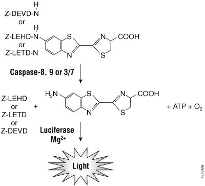
Figure 4. Caspase-8, -9, -3/7 cleavage of the proluminogenic substrates containing LETD, LEHD or DEVD, respectively. Following caspase cleavage, a substrate for luciferase (aminoluciferin) is released, resulting in the production of light.
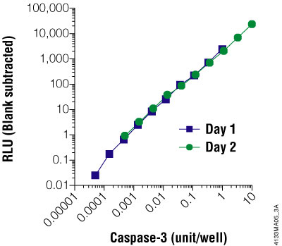
Figure 5. The Caspase-Glo® 3/7 Assay is linear over four orders of magnitude of caspase concentration. Purified caspase-3 was titrated and assayed in 96-well plates using the Caspase-Glo® 3/7 Assay on two different days. Luminescence was measured 1 hour after adding the Caspase-Glo® Reagent to the cells. The graph shows that the assay is linear over four orders of magnitude of caspase concentration. One unit of caspase = 0.07ng protein = 1pmol of substrate (Ac-DEVD-pNA) hydrolyzed/minute per the manufacturer's unit definition. Each point represents the average of four wells. The no-caspase control was subtracted from each point.
The Apo-ONE® Homogeneous Caspase 3/7 Assay detects caspase-3/7 activity via cleavage of a pro-fluorescent DEVD-rhodamine 110 substrate [(Z-DEVD)₂-R110] (Figure 6). The reagent permeabilizes cells, delivers substrate, and stabilizes caspase activity in a single 1:1 addition. Because the fluorescent R110 product accumulates in the presence of active caspase, extending incubation up to 18 hours increases the signal-to-background ratio. The assay is easily scaled for HTS as long as the 1:1 ratio is maintained.
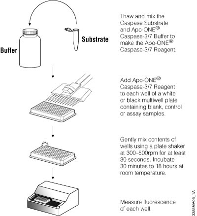
Figure 6. Schematic of Apo-ONE® Assay protocol.
During apoptosis the electrochemical gradient across the MOM collapses. A common method uses a lipophilic cationic dye (e.g., JC-1): in healthy cells the dye accumulates in the mitochondria and forms aggregates emitting bright red fluorescence, whereas in apoptotic cells the dye diffuses into the cytoplasm where the monomeric form emits green fluorescence (Zamzami et al. 2000). Other dyes report redox potential or metabolic activity. Because mitochondrial dyes cannot by themselves distinguish apoptosis from necrosis, they are usually used together with a caspase assay (Waterhouse et al. 2003).
Eukaryotic cells normally maintain phospholipid asymmetry; during early-to-mid cell death, PS becomes abundant on the outer leaflet. Annexin V, a phospholipid-binding protein with high affinity for PS, does not bind intact cells but binds dying cells. Conjugated to a dye or fluorophore, Annexin V can label apoptotic cells for detection by microscopy or flow cytometry (van Genderen et al. 2003; Bossy-Wetzel and Green, 2000).
Annexin V-based detection can be implemented in multiple formats, ranging from endpoint assays to real-time kinetic measurement. Live-cell, non-lytic approaches are particularly valuable, they provide information on the timing of events and keep cells intact for additional use. For example, take the RealTime-Glo™ Annexin V Apoptosis and Necrosis Assay. This assay employs simultaneous detection of PS exposure (via luminescent Annexin V fused to NanoBiT® luciferase) and secondary necrosis (via pro-fluorescent DNA dye) allows accurate determination of apoptotic onset in a single well. Because cells remain intact throughout measurement, the same plate can be read repeatedly, eliminating the need for multiple time-course plates. This approach is well-tolerated by many cultured cell types and supports same-well multiplexing with orthogonal assays.
In apoptotic cells, genomic DNA is cleaved into multimers of 180–200bp, observable as a "ladder" by gel electrophoresis. To detect fragmentation at the single-cell level, the DeadEnd™ Assays use the TUNEL (TdT-mediated dUTP Nick End Labeling) approach: after permeabilization, the 3′ OH ends of the fragments are labeled with fluorescein-12-dUTP (fluorometric) or biotinylated nucleotides detected with streptavidin-HRP (colorimetric).
How Do You Induce Apoptosis in Cells?
Apoptosis can be induced by protein synthesis inhibitors (anisomycin) or DNA topoisomerase I inhibitors (camptothecin) in HL-60 cells; by protein kinase inhibitors (staurosporine); by growth factor withdrawal (e.g., NGF-deprivation of PC12 cells; Batistatou and Greene, 1991); by glucocorticoids (dexamethasone) in mouse thymus lymphocytes; and by activation of Fas or TNF receptors with the respective ligands or agonist antibodies (Tewari and Dixit 1995). Three worked induction systems are commonly used:
How Do You Choose the Right Apoptosis Assay?
Choose your apoptosis assay based on three factors: the apoptotic stage you're measuring (early vs. late), your detection format preference (kinetic real-time vs. endpoint), and your experimental model (2D vs. 3D). Note that kinetic measurement in 3D may require specialized plate readers or imaging systems—verify compatibility with your equipment and consult each product's technical manual for validated conditions.
Why Study Apoptosis in 3D Cell Culture Models?
3D cell culture models (spheroids, organoids, and organs-on-a-chip scaffolds) provide physiologically relevant models to collect apoptosis data. In 3D environments, cells experience realistic gradients of nutrients, oxygen, and signaling molecules, leading to complex and ideally more realistic apoptotic responses not captured in 2D monolayers. Increased federal regulation around New Approach Methodologies (NAMs) has led to more toxicology researchers using these models to test drug responses. 3D model readouts correlate well with clinical outcomes of cancer research, raising the value of measuring apoptosis directly in these models (Xiang et al. 2024).
Key advantages of studying apoptosis in 3D culture include:
- Better recapitulation of in vivo apoptotic patterns, including hypoxia-driven apoptosis in spheroid cores.
- Enhanced predictivity of compound efficacy and toxicity.
- Ability to observe cell–cell communication effects on apoptosis.
How Do You Apply Multiplexing to Apoptosis Assays?
Multiplexing—gathering more than one dataset from the same sample—has many benefits. It is extremely useful for generating internal controls and following two biological processes without requiring repeating work (Figures 7 and 8). The protocols below are starting points and require optimization for each experimental system; run appropriate controls, including each assay individually. Detailed background and recommended controls accompany each product.
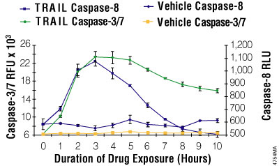
Figure 7. Multiplexing luminescent caspase-8 and fluorescent caspase-3/7 assays. Jurkat cells were seeded at 25,000 cells/well. Fifty microliters of rTRAIL (Chemicon, 100ng/ml final) or a vehicle control (RPMI 1640 with 10% FBS) was added to replicate wells every hour for 10 hours. Caspase-Glo® 8 reagent was prepared by combining the assay buffer with the substrate. The fluorescent Apo-ONE® Assay caspase-3/7 substrate was mixed into the Caspase-Glo® 8 reagent at a final concentration of 50μM. The combined reagent/substrate was added in 100μl volumes, incubated 60 minutes, and then luminescence and fluorescence were measured.
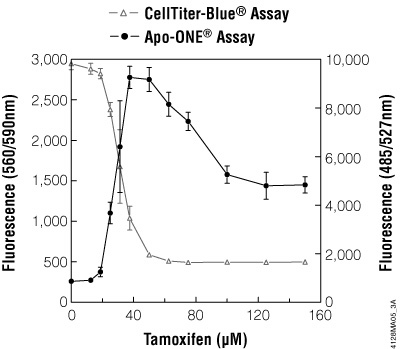
Figure 8. Multiplexing cell viability assays. HepG2 cells (10,000 cells/100μl cultured overnight) were treated with various concentrations of tamoxifen for 5 hours. Viability was determined by adding CellTiter-Blue® Reagent (20μl/well) to each well after 3.5 hours of drug treatment and incubating for 1 hour before recording fluorescence (560Ex/590Em). Caspase activity was then determined by adding 120μl/well of Apo-ONE® Reagent and incubating for 0.5 hour before recording fluorescence (485Ex/527Em).
Combine a luminescent Caspase-Glo® 8 or 9 reagent with the fluorescent Apo-ONE® caspase-3/7 substrate (1:200) in one reagent; read luminescence then fluorescence. Distinguishes extrinsic (caspase-8) vs. intrinsic (caspase-9) initiation.
Learn more: Caspase-Glo® 8 Assay, Caspase-Glo® 9 Assay, Apo-ONE® Homogeneous Caspase-3/7 Assay.
Add CellTiter-Blue® during the final 1–2 h of treatment (read 560/590), then Apo-ONE® Reagent (read 485/527).
Learn more: CellTiter-Blue® Cell Viability Assay, Apo-ONE® Homogeneous Caspase-3/7 Assay.
Read CellTiter-Blue® fluorescence (560/590), then add Caspase-Glo® 3/7 Reagent and read luminescence. No spectral overlap.
Learn more: CellTiter-Blue® Cell Viability Assay, Caspase-Glo® 3/7 Assay.
Pair CytoTox-ONE™ membrane integrity (necrosis; read 560/590) with Caspase-Glo® 3/7 (apoptosis; luminescence).
Learn more: CytoTox-ONE™ Homogeneous Membrane Integrity Assay, Caspase-Glo® 3/7 Assay.
Use EnduRen™ Live Cell Substrate with a Renilla luciferase reporter (read luminescence), then Apo-ONE® Reagent (read 485/527).
Learn more: EnduRen™ Live Cell Substrate, Apo-ONE® Homogeneous Caspase-3/7 Assay Reagent.
Frequently Asked Questions
Q1: What is the difference between apoptosis and necrosis?
A: Apoptosis is programmed, controlled cell death with minimal inflammatory response. It is essential for normal development and homeostasis, but excessive apoptosis can be linked to disease states. Necrosis is classically uncontrolled, traumatic cell death that damages surrounding tissue and triggers inflammation. Some necrosis can be regulated (necroptosis).
Q2: Why is more than one method needed to confirm apoptosis?
A: Apoptosis markers can be transient, and apoptotic cells in vitro eventually undergo secondary necrosis. A single marker may miss the mechanism or overlap with necrosis, so combining methods (e.g., a caspase assay plus DNA fragmentation or annexin V) and understanding your model's kinetics gives a more reliable determination.
Q3: Which assay distinguishes the extrinsic from the intrinsic pathway?
A: Caspase-8 activity is associated with the extrinsic (death receptor) pathway and caspase-9 with the intrinsic (mitochondrial) pathway; Caspase-Glo® 8 and 9 measure these, and multiplexing with a Caspase-Glo®-3/7 assay confirms effector activation.
Q4: Can Promega apoptosis assays be used in 3D cultures or spheroids?
A: Promega has a broad range of assays designed for the detection of apoptosis in 3D models:
- Caspase-Glo® 3/7 3D Assay: same chemistry as the classic Caspase-Glo® 3/7 Assay, with a protocol validated across scaffold-free (ULA-plate spheroid) and scaffold-based (Matrigel®-embedded) models.
- RealTime-Glo™ Annexin V Apoptosis and Necrosis Assay: preliminary data support real-time PS-exposure measurement in spheroid models; well suited to formats compatible with kinetic reads.
- Multiplex options in 3D: CellTox™ Green Cytotoxicity Assay: Multiplex option for membrane integrity, designed for extended 3D exposures; CellTiter-Glo® 3D Cell Viability Assay: Multiple option. ATP-based viability optimized for penetration into microtissues.
Q5: How do I know my signal is caspase-specific?
A: Include a positive control (a known inducer such as anti-Fas, staurosporine or anisomycin), a negative/vehicle control, and a caspase-inhibitor control that should reduce the signal.
Q6: At what apoptosis stage should I measure to detect a compound's effect?
A: This choice depends on your specific goal. When detecting whether a compound triggers apoptosis at all, effector caspase-3/7 activity gives the most robust, sensitive signal because both pathways converge there. Early-stage markers (Annexin V, mitochondrial potential) catch the event sooner, but the early-apoptotic window is brief, so detection depends more on assay timing than with endpoint markers. Late-stage readouts (membrane permeabilization) confirm end-stage death but overlap with secondary necrosis.
Q7: Which assay should I use for high-throughput screening?
A: Luminescent assays are best suited for high-throughput screening due to their sensitivity and add-mix-measure protocols. Our Caspase-Glo® 8 and Caspase-Glo® 9, assays are configured for 384-well formats. Our Caspase-Glo® 3/7 and Caspase-Glo® 3/7 3D assays are configured for both 384- and 1536-well formats. and 3/7 3D assays.
Q8: Can I use the same assay for 2D and 3D cultures?
A: It depends. Many of our assays are compatible with both 2D and 3D formats. However, you may need to optimize lysis buffers, incubation times, or sampling protocols for spheroids. Likewise, we also offer 3D-specific versions of select assays, such as the Caspase-Glo® 3/7 3D Assay. Contact technical support for 3D application guidance.
Q9: What is the typical time-to-result for apoptosis assays?
A: Endpoint assays generally give results within a few hours of the read step, while real-time (kinetic) assays such as RealTime-Glo™ Annexin V allow continuous reading of the same sample over time, revealing apoptotic onset earlier than single-endpoint methods. Actual timing depends on the cell model and inducer.
Q10: What is annexin V and how is it used to detect apoptosis?
A: Annexin V is a protein with a high affinity for PS. In healthy cells, PS sits on the inner membrane leaflet, but during apoptosis it flips to the outer leaflet, where labeled annexin V binds it. Promega’s RealTime-Glo™ Annexin V Assay detects this binding in real time.
Q11: How do you distinguish apoptosis from necrosis experimentally?
A: Combine markers that separate the two processes. Caspase activity (e.g., Caspase-Glo® 3/7) indicates apoptosis, while loss of membrane integrity (e.g., CytoTox-ONE™) indicates necrosis. Run both on the same sample: apoptotic cells show caspase activity with intact membranes; necrotic cells show membrane damage with little caspase activity.
References
- Batistatou, A. and Greene, L.A. (1991) Aurintricarboxylic acid rescues PC12 cells and sympathetic neurons from cell death caused by nerve growth factor deprivation: Correlation with suppression of endonuclease activity. J. Cell Biol. 115, 461–71.
- Boatright, K.M. et al. (2003) A unified model for apical caspase activation. Molecular Cell 11, 529–41.
- Bossy-Wetzel, E. and Green, D.R. (2000) Detection of apoptosis: Annexin V labeling. In: Meth. Enzymol. Reed, J.C. ed. 332, 15–18.
- Chen, M., Wang, L., Li, M., Budai, M. M., & Wang, J. (2022). Mitochondrion-mediated cell death through Erk1-Alox5 independent of caspase-9 signaling. Cells, 11 (19), 3053. https://doi.org/10.3390/cells11193053
- Cotter, T.G. and Curtin, J.F. (2003) Historical perspectives. In: Essays in Biochemistry. Cotter, T.G. et al. eds. 39, 1–10.
- Galluzzi, L., et al. (2024). Targeting immunogenic cell stress and death for cancer therapy. Nature reviews Drug discovery, 23 (6), 445-460.
- Gupta, G., Samuel, V.P., Rekha, M.M., Rani, B., Sasikumar, Y., Nayak, P.P., Sudan, P., Goyal, K., Oliver, B.G., Chakraborty, A., & Dua, K. (2025). Caspase-independent cell death in lung cancer: from mechanisms to clinical applications. Naunyn-Schmiedeberg's Archives of Pharmacology. https://doi.org/10.1007/s00210-025-04149-0
- Hengartner, M.O. (1997) Programmed Cell Death. In: C. elegans II. Riddle, D.L. et al. eds. 383–496. (Cold Spring Harbor Laboratory Press, Plainview, N.Y.)
- Hengartner, M.O. (2000) The biochemistry of apoptosis. Nature 407, 770–6.
- Kaloni, D., et al. (2023). BCL-2 protein family: attractive targets for cancer therapy. Apoptosis, 28 (1), 20-38.
- Kari, S., et al. (2022). Programmed cell death detection methods: a systematic review and a categorical comparison. Apoptosis, 27 (7), 482-508.
- Kerr, J.F.R., et al. (1972) Apoptosis: A basic biological phenomenon with wide-ranging implications in tissue kinetics. Br. J. Cancer 26, 239–57.
- Leist, M. and Jäättelä, M. (2001) Four deaths and a funeral: From caspases to alternative mechanisms. Nat. Rev. Mol. Cell Biol. 2, 589–98.
- Moyer, A., Tanaka, K., & Cheng, E. H. (2025). Apoptosis in cancer biology and therapy. Annual Review of Pathology: Mechanisms of Disease, 20 (1), 303-328.
- Murphy, F.J., Serry, L.T. and Hayes, I. (2003) Therapeutic approaches to the modulation of apoptosis. In: Essays in Biochemistry. Cotter, T. et al. eds. 39, 131–53. (Cold Spring Harbor Laboratory Press, Plainview, N.Y.)
- Parone, P. et al. (2003) Apoptosis: Bombarding the mitochondria. In: Essays in Biochemistry. Cotter, T. et al. eds. 39, 41–51. (Cold Spring Harbor Laboratory Press, Plainview, N.Y.)
- Pulica, R., et al. (2025). Dys-regulated phosphatidylserine externalization as a cell intrinsic immune escape mechanism in cancer. Cell Communication and Signaling, 23 (1), 131.
- Rajesh & Kanneganti (2022), Innate Immune Cell Death in Neuroinflammation and Alzheimer's Disease, Cells — apoptosis/pyroptosis/necroptosis crosstalk in neurodegeneration (a modern replacement for the Aβ/caspase-12 material). https://doi.org/10.3390/cells11121885
- Rich, T., Watson, C.J. and Wyllie, A. (1999) Apoptosis: The germs of death. Nat.Cell Biol. 1, E69–E71.
- Riss, T.L. and Moravec, R.L. (2004) Use of multiple assay endpoints to investigate the effects of incubation time, dose of toxin, and plating density in cell-based cytotoxicity assays. Assay and Drug Dev.Technol. 2, 51–62.
- Tewari, M. and Dixit, V.M. (1995) Fas- and tumor necrosis factor-induced apoptosis is inhibited by the poxvirus crmA gene product. J. Biol. Chem. 17, 3255–60.
- van Genderen, H. et al. (2003) Membrane alterations in dying cells. In: Cell Proliferation and Apoptosis. Hughes, D and Mehmet, H., eds. 185–200. (Bios Scientific Publishers, Ltd. Oxford)
- Wajant, H. et al. (2003) Death Receptors. In: Essays in Biochemistry. Cotter, T. et al. eds. 39, 53–71. (Cold Spring Harbor Laboratory Press, Plainview, N.Y.)
- Wang, X., Grus, W. E., & Zhang, J. (2006). Gene losses during human origins. PLoS biology, 4(3), e52.
- Waterhouse, N.J. et al. (2003) Mitochondrial outer-membrane permeabilization in apoptosis. In: Cell Proliferation and Apoptosis. Hughes, D. and Mehmet, H., eds. 185–200. (Bios Scientific Publishers, Ltd. Oxford)
- Williamson, P., et al. (2000) Phosphatidylserine exposure and phagocytosis of apoptotic cells. Meth. Cell Biol. 66, 339–64.
- Wyllie, A. H. (1980) Glucocorticoid-induced thymocyte apoptosis is associated with endogenous endonuclease activation. Nature 284, 555–6.
- Xiang, D., et al. (2024). Building consensus on the application of organoid-based drug sensitivity testing in cancer precision medicine and drug development. Theranostics, 14 (8), 3300.
- Zamzami, N. et al. (2000) Quantitation of mitochondrial transmembrane potential in cells and in isolated mitochondria. In: Methods Enzymol. Reed J.C., ed. 322, 208–13.
- Zou, H. et al. (1997) Apaf-1, a human protein homologous to C. elegans CED-4, participates in cytochrome-c dependent activation of caspase-3. Cell 90, 405–13.
Apo-ONE, Caspase-Glo, CellTiter-Blue are CellTiter 96 are trademarks of Promega Corporation and are registered with the U.S. Patent and Trademark Office. CaspACE, CytoTox-ONE, DeadEnd, and EnduRen are trademarks of Promega Corporation.
Cy is a registered trademark of Amersham Biosciences Ltd. FACS is a registered trademark of Becton Dickinson and Company. HemoDe is a registered trademark of Scientific Safety Solvents. FluoroNunc is a trademark of Nalge Nunc International. Lab-Tek is a registered trademark of Nalge Nunc International. NANOpure are registered trademarks of Barnstead/Thermolyne Corporation. Parafilm is a registered trademark of American National Can Company. PolyPrep is a registered trademark of Bio-Rad Laboratories, Inc. Prionex is a registered trademark of Pentapharm Ltd. SlowFade is a registered trademark of Molecular Probes, Inc. Superfrost is a registered trademark of Erie Scientific. Triton is a registered trademark of Union Carbide Chemicals and Plastics Technology Corporation. Tween is a registered trademark of ICI Americas, Inc.VECTASHIELD is a registered trademark of Vector Laboratories, Inc.
Products may be covered by pending or issued patents or may have certain limitations. Please visit our website for more information.
All prices and specifications are subject to change without prior notice.
Product claims are subject to change. Please contact Promega Technical Services or access the Promega online catalog for the most up-to-date information on Promega products.


