Kinase Target Engagement
The NanoBRET® Target Engagement (TE) Intracellular Kinase Assays provide a way to quantitatively measure specific kinase-inhibitor interactions in live cells using Bioluminescence Resonance Energy Transfer (BRET). The assay measures the apparent cellular affinity of test compounds bound by competitive displacement of a cell-permeable fluorescent NanoBRET® tracer, reversibly bound to a kinase-NanoLuc® luciferase fusion in cells.
NanoBRET® Kinase Target Engagement Assays
- Measure Kinase Target Engagement in Live Cells: Quantify compound affinity & fractional occupancy for multiple types of kinase inhibitors (I-IV).
- Assays for over 340 Kinases: Ready-to-use assays span the kinome, readily enabling selectivity analysis.
- Use Full-Length Kinase: Assays use full-length wild-type kinases. Select mutant kinase or domain-specific kinase assays are available.
- Multi-Well Plate Format: Simple assay method is scalable from 96-well to 384-well or beyond.
- Assess Residence Time: Determine duration of test compound binding to target kinase in live cells.
What You Need to Perform a NanoBRET® TE Assay

The tracers and substrate/inhibitor combinations are also available as standalone products.
Filter By
Shop All Kinase Target Engagement Products
Showing 16 of 16 Products
How NanoBRET® Kinase Target Engagement Assays Work
A Kinase-NanoLuc® fusion vector is introduced by transfection into mammalian cells and allowed to express. As NanoLuc® luciferase is extremely bright, only low expression of the kinase-NanoLuc® fusion protein is needed.
In the NanoBRET® TE Kinase Assay, a cell-permeable fluorescent NanoBRET® tracer, a NanoLuc® substrate, and the Extracellular NanoLuc® Inhibitor are supplied. Addition of these to cells expressing the kinase-NanoLuc® fusion allows a strong BRET signal to be achieved between the kinase-NanoLuc® protein and NanoBRET® tracer. The extracellular NanoLuc® inhibitor ensures that the BRET signal achieved is from live, uncompromised cells.
The presence of unlabeled test compound that binds to target kinase results in a loss of BRET signal. As BRET has tight distance constraints, the data obtained is specific for the kinase fused to NanoLuc® luciferase. In addition, the data results in a quantitative intracellular affinity provided the appropriate tracer concentration is used.

Example Kinase Target Engagement Data
Measure Affinity, Selectivity, Potency and Residence Time
Obtain Quantitative Measurement of Compound Intracellular Affinity

Determine Compound Cellular Selectivity and Fractional Occupancy for Related Kinases

NanoBRET® Target Engagement can be used to compare inhibitor affinity for wild-type (WT) and mutant kinases. NanoBRET® TE Intracellular Kinase Assay K-10 was used for JAK and JAK(V617F), which is a clinically acquired mutation found in myeloproliferative cancers. Panel A: Engagement of Type I ATP competitive inhibitors against JAK2(V617F). Panel B: Target engagement potency was stronger for Type I inhibitor ruxolitinib with JAK2(V617F) versus JAK2 wild-type. Panel C: This differed from the finding with the Type II inhibitor CHZ-868, which had similar affinity for both JAK2(V617F) mutant and JAK2 wild-type kinases.
Achieve Improved Correlation between Compound Affinity and Downstream Functional Potency

Assess Compound Residence Time in Live Cells


FAQ
The technical manuals for the NanoBRET® TE Intracellular Kinase Assays (#TM598 and #TM603) utilize HEK293 cells. However, other cell types have been used, such as HeLa and U2OS. If cell types different from HEK293 are used, it's recommended that the transfection conditions to introduce the kinase-NanoLuc® fusion vector are optimized.
The new adherent (ADH) assay format is more efficient for the user compared to the original non-binding surface (NBS) format. The ADH format involves the use of adhered cells in a tissue culture-treated plate.
The NBS format requires that cells are transfected in plates or dishes the day prior to addition to the assay plate. On the day of the assay, the cells are harvested and seeded in non-binding surface assay plates prior to running the assay. Nonbinding surface plates are ideal for certain tracers with suboptimal properties that prevent their use in conventional polystyrene assay plates.
The ADH format uses tissue culture treated plates and allows the user to transfect the kinase-NanoLuc® fusion vector in the assay plate. The harvesting and seeding step is not needed with the ADH format, as the cells are transfected and seeded in the assay plate the day before.
All available kinases and kinase complexes are listed here with links to application notes and data. If the kinase has also been tested in the NBS format, then a separate application note link is provided in the table. Application notes are also available on the product pages for each individual kinase-NanoLuc® fusion vector.
For each kinase, there is one recommended assay/tracer, as it provides the largest assay window. However, several kinases have additional application notes that use alternate assays. Additional data beyond the recommended assay is available on the kinase-NanoLuc® fusion vector product page when available.
Depending on your needs, having an assay with the largest window may be advantageous, so the recommended assay would be the one to select. In other cases, it may be preferable to work with the same assay/tracer for multiple kinases. In this case, the recommended assay may not be the same for each kinase studied. Please refer to #TM598 or #TM603 for guidelines on assay window and assay capabilities.
Additional kinase-NanoLuc® fusion vectors and NanoBRET® TE Kinase Assays have been developed and are available through our Tailored R&D Solutions (TRS) service. If an assay has not been developed for the kinase, the TRS team could develop one for you or guide you to develop your own assay. A resource for developing NanoBRET® TE assays is also available in this Methods in Molecular Biology chapter. Please inquire here.
Publications
Quantitative, Wide-Spectrum Kinase Profiling in Live Cells for Assessing the Effect of Cellular ATP on Target Engagement
In this 2018 Cell Chemical Biology article, Vasta et al. describe a bioluminescence resonance energy transfer (BRET) -based method for measuring kinase target engagement inside live cells.
A High-Throughput BRET Cellular Target Engagement Assay Links Biochemical to Cellular Activity for Bruton's Tyrosine Kinase
In this 2019 SLAS Discovery article, Ong et al. use NanoBRET® TE methods to characterize target occupancy for BTK.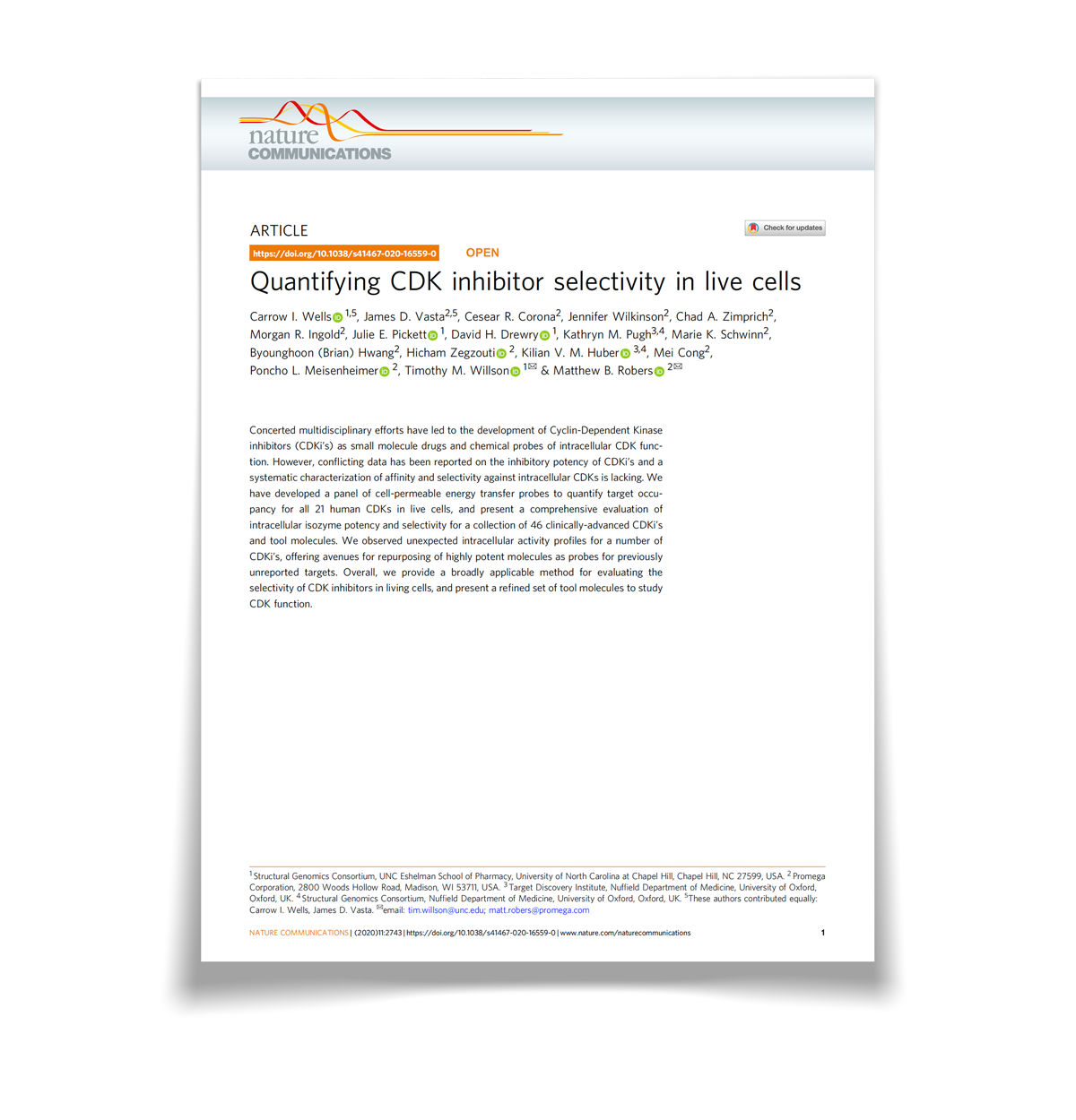
Quantifying CDK Inhibitor Selectivity in Live Cells
In this 2020 Nature Communications paper, teams at the Structural Genomics Consortium and Promega used the NanoBRET® Target Engagement technology to uncover surprising patterns of selectivity for touted CDKIs and abandoned clinical leads.
How Can We Help You?
In addition to the pre-built kinase assays featured on this page, we also offer kits and reagent to build your own NanoBRET® Target Engagement assays, and custom assay development capabilities.