Lumit® Cell Proliferation Assay (Human Ki-67)
No-Wash Assay for hKi-67 Detection
- Increase confidence in your data with a robust assay response
- Decrease prep time using a fast, no-wash, add-and-read protocol
- Make quicker decisions by using an earlier readout of proliferation


Catalog Number:
Size
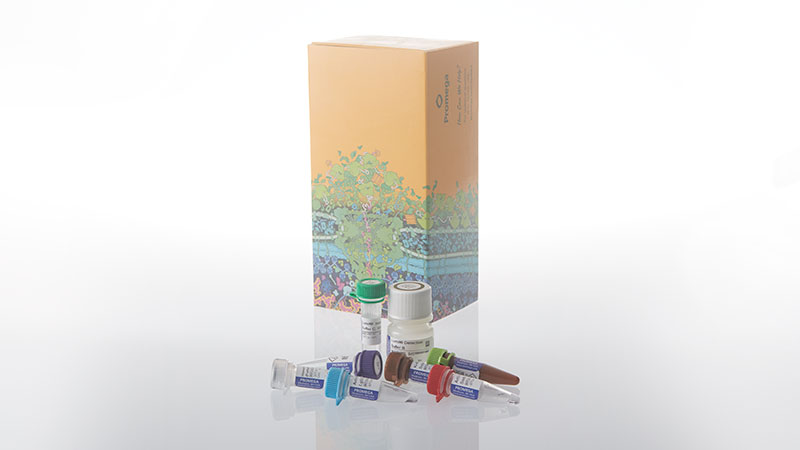
Catalog Number: GC1000
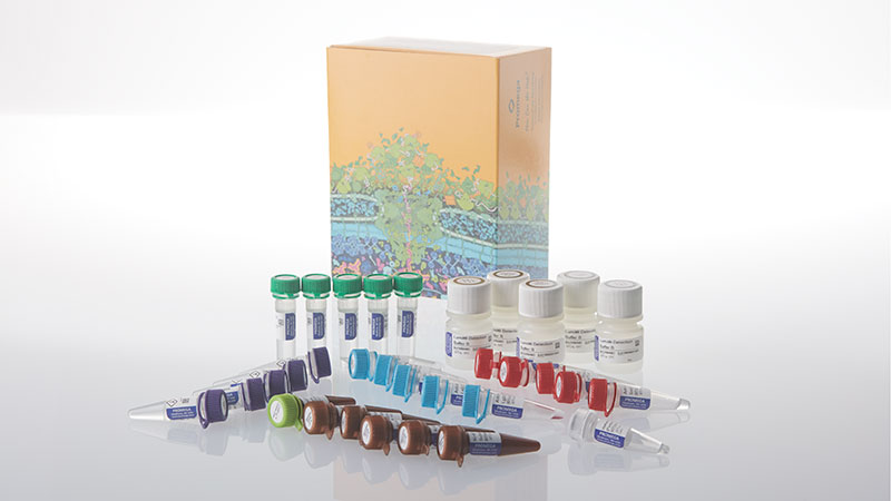
Catalog Number: GC1002
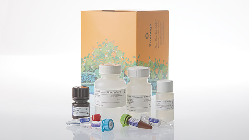
Catalog Number: GC1001
Reliable Proliferation Results in Hours, Not Days
Current methods to assess cell proliferation are laborious, unreliable, and can require treatment times of 72–96 hours — increasing hands-on time, reducing confidence in results, and delaying decision-making.
The Lumit® Cell Proliferation Assay (Human Ki-67) is an add-and-read plate-based assay that enables researchers to track hKi-67, a well-known marker of cell proliferation, without complicated washing steps. The kit includes CellTox™ Green Dye for multiplexed detection of cytotoxicity and proliferation in the same well. With a robust response detectable at earlier time points, researchers can generate more data with higher confidence along with reduced prep work and time to results.

Watch the video to hear how researcher Clara Gouez is using the Lumit® Cell Proliferation Assay (Human Ki-67) to advance her gastric cancer research.
How the Lumit® Cell Proliferation Assay Works
The Lumit® Cell Proliferation Assay (Human Ki-67) is based on Lumit® Technology. Primary antibodies to hKi-67 were selected for their specific and sensitive detection and labeled with the LgBiT and SmBiT subunits of NanoBiT® Luciferase. In the presence of hKi-67, the subunits are brought together to form an active luciferase enzyme. Addition of optimized substrate generates a bright luminescent signal proportional to hKi-67 levels.

Simple Protocol Requires No Wash Steps

Compared with conventional fixation-based Ki-67 flow cytometry, the assay is significantly simpler and faster, requiring no dissociation or wash steps while still producing robust and reproducible results.
Zeynep Kaya, PhD, Postdoctoral Research Fellow, Prof Andrew Beggs Group, University of Birmingham
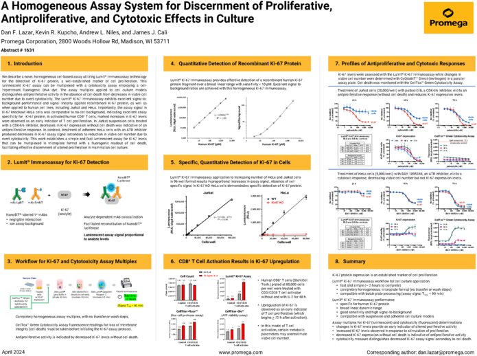
Broad Linear Range and Specific hKi-67 Detection
The Lumit® Cell Proliferation Assay demonstrates excellent linearity in microplate formats and specifically detects hKi-67 levels.


Measure Decreases in Cell Proliferation


Treatment of Jurkat cells (10,000/well, 384-well plate, ¼ standard 96-well volumes) with increasing concentrations of compounds. Both compounds reduced hKi-67 expression levels in a dose-dependent manner; however, BAY-1895344 induced cytotoxicity. Palbociclib produced a large change in hKi-67 levels without causing cytotoxicity after only 24 hours of treatment, enabling earlier assessment of compound effects on proliferation. Palbociclib’s effect appears minimal based on total ATP content.
Measure Increases in Cell Proliferation


Human CD8+ T cells (StemCell Tech) plated at 80,000 cells per well were treated with CD3/CD28 T cell activator with (teal) and without (purple) 10ng/ml IL-2 for 48 hours. Upregulation of hKi-67 is observed with the Lumit® Cell Proliferation Assay before T cell proliferation (which begins >72 hours after activation), demonstrating use of this assay as an early indicator of proliferation.
Compatible with 3D Culture Methods

In 3D spheroids, decreases in hKi-67 protein levels provide a clear and early indication of antiproliferative activity with a larger response window than a metabolic measure of viable cell number. HCT116 cells (1,000 cells/well) were plated in SBio PrimeSurface 96U plates (ULA; round bottom) and grown for 3 days to form 3D spheroids. The resultant HCT116 spheroids were then treated for 24 hours with increasing concentrations of nutlin-3a. Subsequently, proliferation was assessed with the Lumit® Cell Proliferation Assay (Human Ki-67) and a metabolic activity assay in separate plates. Note: Day 4 untreated HCT116 spheroids were ~440µm in diameter.
Protocols
Specifications
Catalog Number:
What's in the box?
| Item | Part # | Size |
|---|---|---|
CellTox™ Green Dye, 1,000X |
G873A | 1 × 20μl |
Human Ki-67 Protein (Partial) Positive Control |
GC100A | 1 × 25μl |
Anti-hKi-67 mAb SmBiT, 400X |
GC101A | 1 × 60μl |
Anti-hKi-67 mAb LgBiT, 400X |
GC102A | 1 × 60μl |
Lumit® Immunoassay Buffer C, 10X |
VB115C | 1 × 1.8ml |
Lumit® Lysis Buffer II, 10X |
VB310C | 1 × 1.3ml |
Ki-67 Assay Substrate |
VB321A | 1 × 600μl |
Lumit® Detection Buffer B |
VB406D | 1 × 6ml |
SDS
Search for SDSCertificate of Analysis
Use Restrictions
For Research Use Only. Not for Use in Diagnostic Procedures.Storage Conditions
U.S. Pat. No. 8,809,529, European Pat. No. 2635582, Japanese Pat. No. 5889910 and other patents and patents pending.
U.S. Pat. Nos. 9,797,889, 9,797,890, 10,107,800 and 11,493,504; European Pat. No. 2970412; Japanese Pat. Nos. 7280842 and 7532562; and other patents and patents pending.
What's in the box?
| Item | Part # | Size |
|---|---|---|
CellTox™ Green Dye, 1,000X |
G873B | 1 × 200μl |
Human Ki-67 Protein (Partial) Positive Control |
GC100A | 1 × 25μl |
Anti-hKi-67 mAb SmBiT, 400X |
GC101A | 5 × 60μl |
Anti-hKi-67 mAb LgBiT, 400X |
GC102A | 5 × 60μl |
Lumit® Immunoassay Buffer C, 10X |
VB115C | 5 × 1.8ml |
Lumit® Lysis Buffer II, 10X |
VB310C | 5 × 1.3ml |
Ki-67 Assay Substrate |
VB321A | 5 × 600μl |
Lumit® Detection Buffer B |
VB406D | 5 × 6ml |
SDS
Search for SDSCertificate of Analysis
Use Restrictions
For Research Use Only. Not for Use in Diagnostic Procedures.Storage Conditions
U.S. Pat. No. 8,809,529, European Pat. No. 2635582, Japanese Pat. No. 5889910 and other patents and patents pending.
U.S. Pat. Nos. 9,797,889, 9,797,890, 10,107,800 and 11,493,504; European Pat. No. 2970412; Japanese Pat. Nos. 7280842 and 7532562; and other patents and patents pending.
What's in the box?
| Item | Part # | Size |
|---|---|---|
CellTox™ Green Dye, 1,000X |
G873B | 1 × 200μl |
Human Ki-67 Protein (Partial) Positive Control |
GC100A | 1 × 25μl |
Anti-hKi-67 mAb SmBiT, 400X |
GC101B | 1 × 600μl |
Anti-hKi-67 mAb LgBiT, 400X |
GC102B | 1 × 600μl |
Lumit® Immunoassay Buffer C, 10X |
VB115D | 1 × 18ml |
Lumit® Lysis Buffer II, 10X |
VB310D | 1 × 13ml |
Ki-67 Assay Substrate |
VB321B | 1 × 6ml |
Lumit® Detection Buffer B |
VB406E | 1 × 60ml |
SDS
Search for SDSCertificate of Analysis
Use Restrictions
For Research Use Only. Not for Use in Diagnostic Procedures.Storage Conditions
U.S. Pat. No. 8,809,529, European Pat. No. 2635582, Japanese Pat. No. 5889910 and other patents and patents pending.
U.S. Pat. Nos. 9,797,889, 9,797,890, 10,107,800 and 11,493,504; European Pat. No. 2970412; Japanese Pat. Nos. 7280842 and 7532562; and other patents and patents pending.
Resources
Featured Resource
Need to differentiate between antiproliferative effects and cell death?
Quickly and easily assess cell proliferation with the Lumit® Cell Proliferation Assay (Human Ki-67), a homogeneous cell-based assay for the detection of hKi-67, a key marker of cell proliferation.
Articles
Posters
- Poster: Homogeneous Bioluminescent Immunoassay for hKi-67: A Simple and Robust Screening Tool for Antiproliferative Agents
- Poster: Novel, Luminescent Ki-67 Immunoassay Assesses T Cell Responses
- Poster: Lumit® Immunoassays: Bioluminescent, Sensitive, and Homogeneous Analyte Detection Using Labeled Antibodies
Compare Products
|
Cell Viability (ATP) |
Cell Viability (ATP) |
3D Cell Viability (ATP) |
Real-Time Viability |
Non-Lytic Viability |
Cell Proliferation |
|
|---|---|---|---|---|---|---|
| Best Use | Routine viability & HTS screening | Established protocols using original formulation | 3D spheroids, organoids, microtissues | Kinetic monitoring over time; downstream multiplexing | Multiplex first step; cells needed for follow-up assays | True proliferation readout independent of metabolism |
| Key Decision Points | ||||||
| Measures | Viability | Viability | Viability (3D) | Viability (kinetic) | Viability | Proliferation |
| Cells alive after assay? | ✗ Lytic | ✗ Lytic | ✗ Lytic | ✓ Non-lytic | ✓ Non-lytic | ✗ Lytic |
| Multiplexing compatible? | LimitedLytic—must be terminal step | LimitedLytic—must be terminal step | LimitedLytic—must be terminal step | ✓ ExcellentNon-lytic; pair with any downstream assay | ✓ ExcellentNon-lytic; pair with Caspase-Glo®, CTG, etc. | ModerateCan multiplex with CellTox™ Green or other fluorescent readouts |
| Real-time monitoring? | ✗ Endpoint | ✗ Endpoint | ✗ Endpoint | ✓ Up to 72hRead same wells repeatedly | ✗ Endpoint | ✗ Endpoint |
| 3D culture compatible? | PartialWorks for small spheroids; use 3D version for dense structures | PartialSame as 2.0 | ✓ OptimizedEnhanced lysis for dense 3D structures | PartialSubstrate must penetrate; best for small/loose 3D models | PartialSubstrate access may be limited in dense 3D | ✓ YesDetects Ki-67 in cell lysates from any culture format |
| Assay Attributes | ||||||
| Assay Principle | ATP quantitation (luciferase/luciferin) | ATP quantitation (luciferase/luciferin) | ATP quantitation (enhanced lysis for 3D) | Metabolic reduction of pro-substrate to luciferase substrate | Live-cell protease activity (GF-AFC cleavage) | Ki-67 immunodetection via NanoBiT® complementation |
| Detection Mode | Luminescence | Luminescence | Luminescence | Luminescence | Fluorescence400Ex / 505Em | Luminescence |
| Reagent Format | Ready-to-use liquid | Buffer + lyophilized substrateRequires reconstitution | Ready-to-use liquid | 2 components(enzyme + substrate) | Single reagent | Antibody mix + detection reagent |
| Time to Result | 10min | 10min | ~30min | ContinuousFirst read: 1–2h after addition | 30min | ~2h |
| Practical Considerations | ||||||
| Plate Formats | 96, 384, 1536 | 96, 384, 1536 | 96, 384 | 96, 384 | 96, 384 | 96, 384 |
| HTS Suitability | ✓ Excellent1536-well capable; fast protocol | ✓ Excellent1536-well capable | ✓ Good | ModerateRequires kinetic reader scheduling | ✓ Good | ✓ Good |
| Sensitivity (96-well) | ~15 cells/well | ~10 cells/well | Spheroid-dependent | <100 cells/well | ~40 cells/well | Cell line-dependent |
Similar Products
CellTiter-Glo® Luminescent Cell Viability Assay
Determine the number of viable cells in culture based on quantitation of ATP present.
G7570, G7571, G7572, G7573
CellTiter-Glo® 2.0 Cell Viability Assay
Updated CellTiter-Glo® Cell Viability Assay with improved reagent stability. Quantifies cell proliferation based on ATP detection.
G9241, G9242, G9243
RealTime-Glo™ MT Cell Viability Assay
A bioluminescent method to kinetically monitor viability in cell culture up to 72 hours.
G9711, G9712, G9713
CellTiter 96® AQueous One Solution Cell Proliferation Assay
A single-reagent (MTS), colorimetric method for determining cell viability.
G3582, G3580, G3581