CellTiter-Glo® Luminescent Cell Viability Assay


A Luminescent Assay to Determine the Number of Viable Cells in Culture
- Measures cell viability by quantifying ATP, a direct marker of metabolically active cells
- Detects as few as 15 cells/well in 384-well format; up to 5 logs of linear range
- Simple add-mix-measure protocol—no cell washing, medium removal or multiple pipetting steps
- Glow-type luminescent signal with >5-hour half-life enables flexible batch processing
- Cited in more than 50,000 peer-reviewed publications—one of the most trusted viability assays in life science research
- Compatible with high-throughput screening (HTS), cytotoxicity and proliferation assays
- Integrates with the MyGlo® Reagent Reader
Catalog Number:
Choose a product
Size
Catalog Number: G7570
Catalog Number: G7571
Catalog Number: G7572
Catalog Number: G7573
What Is the CellTiter-Glo® Assay and How Does It Work?
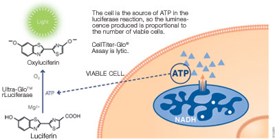
The CellTiter-Glo® Luminescent Cell Viability Assay determines the number of viable cells in culture by quantifying adenosine triphosphate (ATP)—an indicator of metabolically active cells. It uses a bioluminescent luciferase reaction: viable cells release ATP upon lysis, which reacts with the CellTiter-Glo® Reagent to produce a luminescent signal proportional to cell number. The entire protocol requires just one reagent addition and produces results in 10 minutes.
Biochemical Principle: ATP Quantification via Luciferase
ATP is a universal marker of cell viability. Living cells maintain ATP levels through active metabolism, while dead or dying cells rapidly lose ATP. The CellTiter-Glo® Reagent lyses cells on contact, releasing intracellular ATP. The released ATP then drives a luciferase-catalyzed reaction with luciferin, generating bioluminescence that is measured by a compatible luminometer or plate reader.
The luminescence produced is a "glow-type" signal with a half-life generally exceeding 5 hours, depending on cell type and medium. This extended half-life eliminates the need for reagent injectors and allows batch or continuous plate processing—a significant practical advantage in high-throughput environments.
How Do You Perform a CellTiter-Glo® Assay? Key Steps
The assay follows a simple three-step workflow that minimizes hands-on time and potential for error:
- Add CellTiter-Glo® Reagent directly to cells in culture medium (no washing or medium removal required).
- Mix contents for 2 minutes on an orbital shaker to lyse cells.
- Incubate for 10 minutes to stabilize the luminescent signal, then record luminescence.
The homogeneous, "add-mix-measure" format eliminates errors common to multi-step ATP measurement protocols, making CellTiter-Glo® one of the most reproducible viability assays available for multiwell plate formats.

CellTiter-Glo® Assay offers 10-cell sensitivity; up to 5 logs linear range. The signal was read on a MyGlo® Reagent Reader.
Notes:
Using 100μl of CellTiter-Glo® Reagent per assay in a 96-well format, Cat.# G7570 is sufficient to perform 100 assays; Cat.# G7571 and G7572, 1,000 assays; Cat.# G7573, 10,000 assays. Using 25μl of CellTiter-Glo® Reagent per assay in a 384-well format, Cat.# G7570 is sufficient to perform 400 assays; Cat.# G7571 and G7572, 4,000 assays; Cat.# G7573, 40,000 assays.
What Types of Cells and Culture Models Are Compatible with CellTiter-Glo® Assays?
CellTiter-Glo® assays are compatible with virtually all mammalian cell culture models. The lysis-based mechanism works equally well for adherent cells, suspension cultures and more complex 3D model systems.
- Adherent cell lines (e.g., HeLa, A549, MCF-7)
- Suspension cell lines (e.g., Jurkat, K562)
- Primary cells and patient-derived samples
- 3D spheroid and organoid cultures (see the CellTiter-Glo® 3D Assay for optimized 3D performance)
- Cells grown in serum-supplemented or serum-free medium
What Are the Common Applications of the CellTiter-Glo® Assay?
The CellTiter-Glo® Assay is used across a wide range of life science research and drug discovery applications. Its high-throughput compatibility and low coefficient of variation make it a preferred choice for:
- Cell viability and proliferation studies
- Cytotoxicity testing—determining IC50 values for small molecules, biologics, and ADCs
- Drug screening and compound library profiling in 96-, 384-, and 1536-well formats
- Cancer biology—evaluating tumor cell response to chemotherapy, targeted therapy, or radiation
- Antiviral research—quantifying cytopathic effect inhibition (e.g., SARS-CoV-2 antiviral studies)
- Functional genomics—measuring cell survival after RNAi knockdown or CRISPR perturbation
- Immune effector assays—assessing T cell or NK cell cytotoxicity
"We perform knockdown experiments for noncoding RNAs on a number of different cell lines to identify novel cancer therapy targets. The CellTiter-Glo® Assay is an extremely easy-to-use reagent to address cell viability and obtain consistent and reproducible results."
Anan Quan, Research Assistant, Beth Israel Deaconess Medical Center.
"The CellTiter-Glo® Assay provides a very robust, easy-to-use reagent for high-throughput screening of small molecules for antiviral activity against viruses such as SARS-CoV and SARS-CoV-2 that cause a cytopathic effect in cell lines."
Dr. Colleen Jonsson, Director of the Regional Biocontainment Laboratory and Director of the Institute for the Study of Host-Pathogen Systems, University of Tennessee Health Science Center.
Read more about Dr. Jonsson's work to identify antiviral agents.
Customer Testimonials: Advantages of CellTiter-Glo
Pair with the MyGlo® Reagent Reader
Reading your assay results doesn't require a shared plate reader. The MyGlo® Reagent Reader is a compact 96-well luminescence plate reader, enabling you to run luminescent assays at your bench without competing for instrument time. Integrated software generates dose response curves, single concentration and linearity analysis in minutes.

Protocols
Frequently Asked Questions
What are the main differences between the CellTiter-Glo® and CellTiter-Glo® 2.0 Assays?
The CellTiter-Glo® 2.0 Assay is an upgraded version of the original assay, offering a simplified single-reagent format and improved stability. It is particularly suited for high-throughput and automated workflows. The table below summarizes the key differences to help you choose the right version for your application.
| Feature | CellTiter-Glo® (Original) Assay | CellTiter-Glo® 2.0 Assay |
|---|---|---|
| Reagent format | Two-component: lyophilized substrate + buffer (requires reconstitution) | Single, ready-to-use solution (no reconstitution required) |
| Preparation time | ~30min (substrate reconstitution + equilibration) | ~15min (equilibration only) |
| Reagent stability (4°C) | Reconstituted: 48h (~5% loss); 4d (~20% loss) | Stable for repeated use over multiple days at 4°C |
| Sensitivity | Detects as few as 15 cells/well (384-well format) | Detects as few as 15 cells/well (384-well format) |
| Linear dynamic range | Up to 5 logs | Up to 5 logs |
| Signal type | Glow-type; >5h half-life | Glow-type; >5h half-life |
| HTS/automation | Yes: compatible with 96/384/1536-well formats | Yes: optimized for repeated batch use and automation |
| Multiplex compatibility | Standard | Improved; recommended for multiplex workflows |
| Best use case | Standard single-run viability and cytotoxicity assays | High-throughput, multi-day screening campaigns, automation |
When should I choose the CellTiter-Glo® 2.0 Assay over the original?
The CellTiter-Glo® 2.0 Assay is recommended when:
- Your workflow involves automated liquid handling or high-throughput screening platforms requiring a single ready-to-use reagent
- Improved room-temperature reagent stability is important for batch preparation or extended screening campaigns
- You want to minimize preparation variability by eliminating a substrate reconstitution step
The original CellTiter-Glo® Assay remains the preferred choice for standard viability and cytotoxicity assays across conventional plate formats where the reconstituted two-component system is not a limitation.
What controls do I need for a CellTiter-Glo® experiment?
Proper experimental design is critical for reliable CellTiter-Glo® data. Include the following controls on every assay plate:
- Blank control (reagent only, no cells): establishes background luminescence baseline.
- Negative control (cells + vehicle/DMSO at tested concentration): defines 100% viability.
- Positive control (cells + known cytotoxic compound): confirms assay sensitivity and reagent activity.
- Reference standard (cells at defined density): validates signal linearity and enables plate-to-plate normalization.
For dose-response cytotoxicity experiments, use at least 8–10 compound concentrations spanning 3–4 logs to accurately determine IC50 values. Maintain consistent cell seeding density across plates to minimize well-to-well variation.
How does the CellTiter-Glo® Assay detect ATP in cells?
The CellTiter-Glo® Assay lyses cells on contact, releasing intracellular ATP. The released ATP reacts with the luciferase enzyme and luciferin substrate included in the CellTiter-Glo® Reagent, generating bioluminescence that is proportional to the amount of ATP—and therefore to the number of viable cells.
How sensitive is the CellTiter-Glo® Assay compared to other viability assays?
The CellTiter-Glo® Assay detects as few as 15 cells/well in 384-well format, with a linear dynamic range spanning up to 5 logs. This sensitivity significantly exceeds colorimetric assays such as MTT (typically 1,000–5,000 cells minimum) and most fluorescent methods, making the CellTiter-Glo® Assay the preferred choice for miniaturized and high-throughput formats.
Can the CellTiter-Glo® Assay be used with 3D cell cultures or spheroids?
The standard CellTiter-Glo® formulation is not optimized for 3D cultures. Promega offers the CellTiter-Glo® 3D Cell Viability Assay, which contains a modified lysis buffer designed to penetrate and disrupt 3D spheroid structures for accurate viability measurement in complex 3D tumor models.
How long does the CellTiter-Glo® luminescent signal last?
The CellTiter-Glo® glow-type signal has a half-life generally exceeding 5 hours, depending on cell type and medium. This extended stability allows flexible batch processing of multiple plates without requiring reagent injectors or time-sensitive measurement windows.
Is the CellTiter-Glo® Assay compatible with automated liquid handling and HTS platforms?
Yes. The CellTiter-Glo® Assay is designed for multiwell formats (96-, 384-, and 1536-well plates) and is fully compatible with automated liquid handlers, robotic screening platforms, and the Promega MyGlo® Reagent Reader. Its homogeneous format—a single reagent addition with no washing steps—is optimized for high-throughput screening workflows.
What are the limitations of the CellTiter-Glo® Assay?
While the CellTiter-Glo® Assay is highly versatile, understanding its limitations supports better experimental design:
- Endpoint assay only—The CellTiter-Glo® lyses cells and cannot be used for real-time or kinetic viability monitoring within the same wells. For continuous live-cell monitoring, consider RealTime-Glo® MT Cell Viability Assay.
- ATP variability—Intracellular ATP levels can be affected by metabolic state, serum concentration and cell cycle phase. Compounds that alter cellular metabolism (e.g., mitochondrial uncouplers) may produce results that do not accurately reflect cell number.
- Signal saturation—At very high cell densities, signal may plateau. Optimize cell seeding density within the linear range of the assay (typically 500–50,000 cells/well in 96-well format).
- Interference from culture components—Certain culture additives or compound solvents at high concentrations may inhibit luciferase activity. Test solvent controls at equivalent concentrations.
Best practice: Always run a cell number titration in your specific cell line and medium before performing dose-response experiments to confirm linearity and identify the optimal seeding density.
Specifications
Catalog Number:
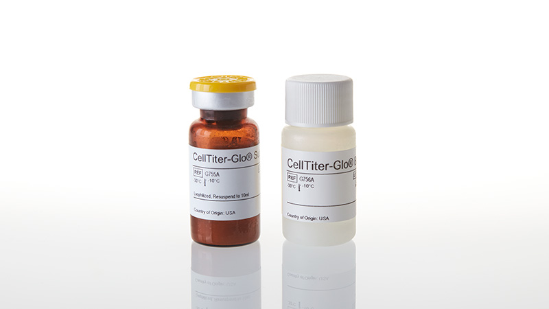
What's in the box?
| Item | Part # | Size |
|---|---|---|
|
CellTiter-Glo® Substrate (lyophilized) |
G755A | 1 × 1 vial |
|
CellTiter-Glo® Buffer |
G756A | 1 × 10ml |
SDS
Search for SDSCertificate of Analysis
Use Restrictions
For Research Use Only. Not for Use in Diagnostic Procedures.Storage Conditions
Reconstituted CellTiter-Glo® Reagent can also be stored at 4°C for 48 hours with ~5% loss of activity or at 4°C for 4 days with ~20% loss of activity.
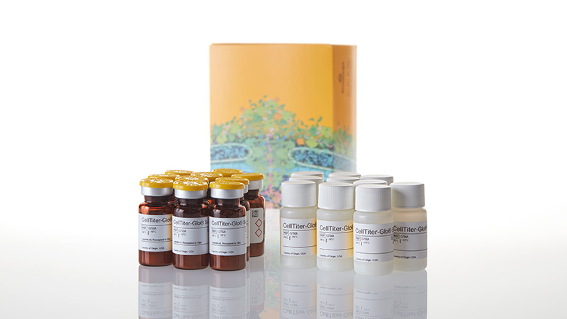
What's in the box?
| Item | Part # | Size |
|---|---|---|
|
CellTiter-Glo® Substrate (lyophilized) |
G755A | 10 × 1 vial |
|
CellTiter-Glo® Buffer |
G756A | 10 × 10ml |
SDS
Search for SDSCertificate of Analysis
Use Restrictions
For Research Use Only. Not for Use in Diagnostic Procedures.Storage Conditions
Reconstituted CellTiter-Glo® Reagent can also be stored at 4°C for 48 hours with ~5% loss of activity or at 4°C for 4 days with ~20% loss of activity.
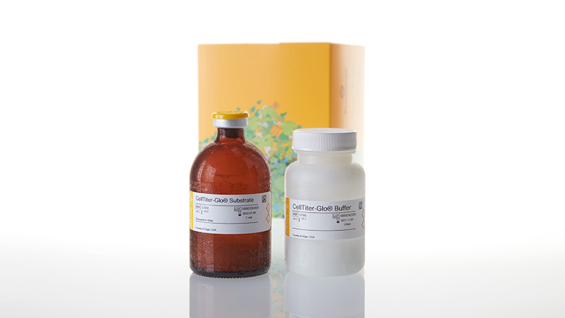
What's in the box?
| Item | Part # | Size |
|---|---|---|
|
CellTiter-Glo® Substrate (lyophilized) |
G755B | 1 × 1 vial |
|
CellTiter-Glo® Buffer |
G756B | 1 × 100ml |
SDS
Search for SDSCertificate of Analysis
Use Restrictions
For Research Use Only. Not for Use in Diagnostic Procedures.Storage Conditions
Reconstituted CellTiter-Glo® Reagent can also be stored at 4°C for 48 hours with ~5% loss of activity or at 4°C for 4 days with ~20% loss of activity.
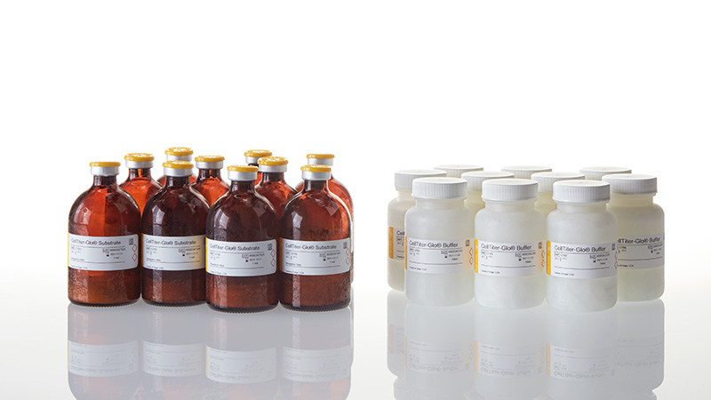
What's in the box?
| Item | Part # | Size |
|---|---|---|
|
CellTiter-Glo® Substrate (lyophilized) |
G755B | 10 × 1 vial |
|
CellTiter-Glo® Buffer |
G756B | 10 × 100ml |
SDS
Search for SDSCertificate of Analysis
Use Restrictions
For Research Use Only. Not for Use in Diagnostic Procedures.Storage Conditions
Reconstituted CellTiter-Glo® Reagent can also be stored at 4°C for 48 hours with ~5% loss of activity or at 4°C for 4 days with ~20% loss of activity.
Resources
Articles
- Engineered virus-like particles for efficient in vivo delivery of therapeutic proteins
- Impact of aerosols on liver xenobiotic metabolism: A comparison of two methods of exposure
- Tolerance to colibactin correlates with homologous recombination proficiency and resistance to irinotecan in colorectal cancer cells
- Compatibility of the Pierce BCA Protein Assay with Promega Lysis Buffers and Lytic Assay Reagents
- Comparative characterization of different molecular formats of bispecific antibodies targeting EGFR and PD-L1
- Overcoming preclinical safety obstacles to discover GDC-2394: A potent and selective NLRP3 inhibitor
- Elevated expression of miR-494-3p is associated with resistance to osimertinib in EGFR T790M-positive non-small cell lung cancer
- Insights into the modular design of kinase inhibitors and application to Abl and Axl
- Metformin attenuates rotenone-induced oxidative stress and mitochondrial damage via the AKT/Nrf2 pathway
- Endogenous retroviruses mediate transcriptional rewiring in response to oncogenic signaling in colorectal cancer
- A CACTA-like transposon in the Anthocyanidin synthase 1 (Ans-1) gene is responsible for apricot fruit colour in the raspberry (Rubus idaeus) cultivar "Varnes"
Other Resources
Compare Products
|
Cell Viability (ATP) |
Cell Viability (ATP) |
3D Cell Viability (ATP) |
Real-Time Viability |
Non-Lytic Viability |
Cell Proliferation |
|
|---|---|---|---|---|---|---|
| Best Use | Routine viability & HTS screening | Established protocols using original formulation | 3D spheroids, organoids, microtissues | Kinetic monitoring over time; downstream multiplexing | Multiplex first step; cells needed for follow-up assays | True proliferation readout independent of metabolism |
| Key Decision Points | ||||||
| Measures | Viability | Viability | Viability (3D) | Viability (kinetic) | Viability | Proliferation |
| Cells alive after assay? | ✗ Lytic | ✗ Lytic | ✗ Lytic | ✓ Non-lytic | ✓ Non-lytic | ✗ Lytic |
| Multiplexing compatible? | LimitedLytic—must be terminal step | LimitedLytic—must be terminal step | LimitedLytic—must be terminal step | ✓ ExcellentNon-lytic; pair with any downstream assay | ✓ ExcellentNon-lytic; pair with Caspase-Glo®, CTG, etc. | ModerateCan multiplex with CellTox™ Green or other fluorescent readouts |
| Real-time monitoring? | ✗ Endpoint | ✗ Endpoint | ✗ Endpoint | ✓ Up to 72hRead same wells repeatedly | ✗ Endpoint | ✗ Endpoint |
| 3D culture compatible? | PartialWorks for small spheroids; use 3D version for dense structures | PartialSame as 2.0 | ✓ OptimizedEnhanced lysis for dense 3D structures | PartialSubstrate must penetrate; best for small/loose 3D models | PartialSubstrate access may be limited in dense 3D | ✓ YesDetects Ki-67 in cell lysates from any culture format |
| Assay Attributes | ||||||
| Assay Principle | ATP quantitation (luciferase/luciferin) | ATP quantitation (luciferase/luciferin) | ATP quantitation (enhanced lysis for 3D) | Metabolic reduction of pro-substrate to luciferase substrate | Live-cell protease activity (GF-AFC cleavage) | Ki-67 immunodetection via NanoBiT® complementation |
| Detection Mode | Luminescence | Luminescence | Luminescence | Luminescence | Fluorescence400Ex / 505Em | Luminescence |
| Reagent Format | Ready-to-use liquid | Buffer + lyophilized substrateRequires reconstitution | Ready-to-use liquid | 2 components(enzyme + substrate) | Single reagent | Antibody mix + detection reagent |
| Time to Result | 10min | 10min | ~30min | ContinuousFirst read: 1–2h after addition | 30min | ~2h |
| Practical Considerations | ||||||
| Plate Formats | 96, 384, 1536 | 96, 384, 1536 | 96, 384 | 96, 384 | 96, 384 | 96, 384 |
| HTS Suitability | ✓ Excellent1536-well capable; fast protocol | ✓ Excellent1536-well capable | ✓ Good | ModerateRequires kinetic reader scheduling | ✓ Good | ✓ Good |
| Sensitivity (96-well) | ~15 cells/well | ~10 cells/well | Spheroid-dependent | <100 cells/well | ~40 cells/well | Cell line-dependent |
Similar Products
CellTiter-Glo® 2.0 Cell Viability Assay
Updated CellTiter-Glo® Cell Viability Assay with improved reagent stability. Quantifies cell proliferation based on ATP detection.
G9241, G9242, G9243
CellTiter-Glo® One Solution Assay
Gold standard method for determining cell viability based on quantitation of ATP.
G8461, G8462
CellTiter-Glo® 3D Cell Viability Assay
A homogeneous method optimized to assess viability in 3D cell culture.
G9681, G9682, G9683
Lumit® Cell Proliferation Assay (Human Ki-67)
Easy add-mix-measure plate-based assay for tracking hKi-67 without washing steps or specialized detection equipment.
GC1000, GC1002, GC1001
Caspase-Glo® 3/7 Assay System
An easy-to-use, plate-based luminescent assay for detecting caspase-3/7 activity.
G8090, G8091, G8093, G8092
Caspase-Glo® 8 Assay Systems
Measure caspase-8 activity with this homogeneous, luminescent assay.
G8200, G8201, G8202
Caspase-Glo® 9 Assay Systems
Measure caspase-9 activity with this homogeneous, luminescent assay.
G8210, G8211, G8212
Frequently Used With
CellTiter-Blue® Cell Viability Assay
Monitor cell viability with this homogeneous, resazurin, fluorescent assay.
G8080, G8081, G8082
BacTiter-Glo™ Microbial Cell Viability Assay
Measure the number of viable microbial cells in culture with a luminescent signal proportional to the amount of ATP present.
G8230, G8231, G8232, G8233
GloMax® Discover System
High-performance microplate reader for detecting luminescence, fluorescence and absorbance.
GM3000
Product Citations
Recent publications that mention the use of this product.