Introduction to 3D Cell Culture
3D cell cultures are increasingly being used for basic research and drug discovery. Here you'll learn what 3D cell cultures are, why and how they are being used, and how to choose assays used with 3D models. Learn about tools for monitoring biology in 3D culture.
This guide provides an overview of 3D cultures and assays. Visit the 3D Assays page to view ordering information for a full list of assays optimized for use in 3D cultures.
What is 3D Cell Culture?
3D cell culture is a culture environment that allows cells to grow and interact with surrounding extracellular framework in three dimensions. This is in contrast with traditional 2D cell cultures in which cells are grown in a flat monolayer on a plate. 3D cell cultures can be grown with or without a supporting scaffold.
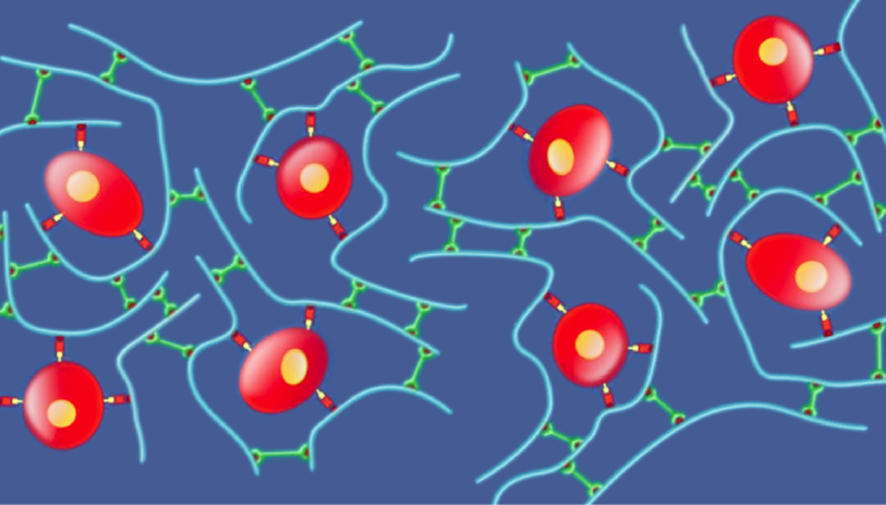
Scaffold 3D Cell Culture
3D cells may be cultured within a supporting scaffold to allow growth in all directions.
Popular types of scaffold include:
Hydrogels: Polymeric material containing a network of crosslinked polymer chains that can absorb and retain water. Hydrogels can be derived from animals (Matrigel®, collagen) or plants (alginate/agarose), or synthesized from chemicals (QGel® Matrix).
Inert matrices: Sponge-like membranes made of polystyrene which contain pores for cells to proliferate and grow.
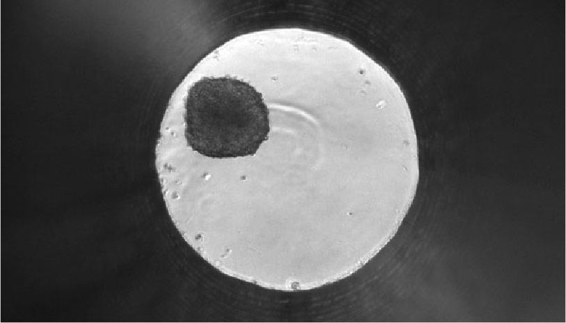
Scaffold-Free 3D Cell Culture
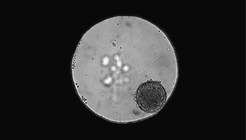
3D cultures can be grown without the presence of a supporting scaffold. Scaffold-free methods rely on cells to self-assemble into clusters or spheroids.
Popular scaffold-free methods include:
Low-adhesion plates: Plates are coated with hydrophilic polymer to prevent cells from sticking to surface, allowing cells to cluster together and form their own extracellular matrix (ECM).
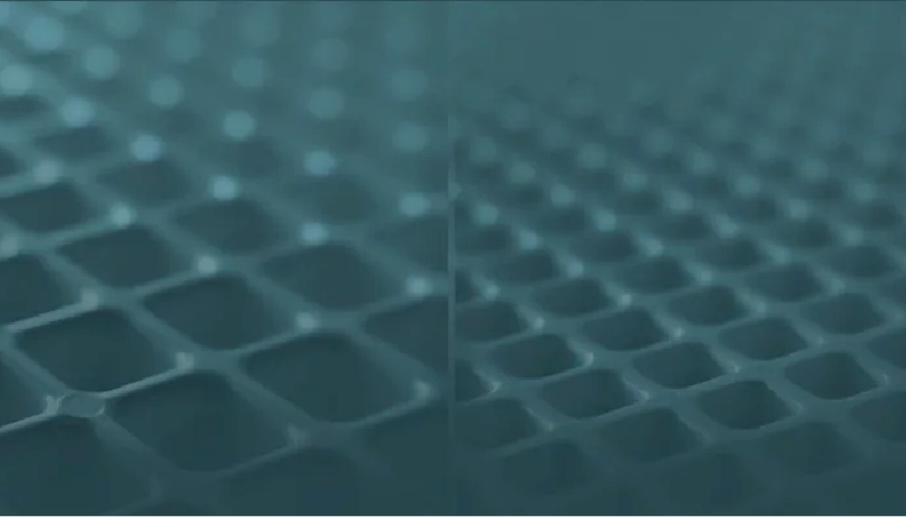
Micropatterned surfaces: Plastic surfaces are modified to provide a micropattern or multiwells, which induce cells to grow as clusters.
Hanging drop: Cells are placed in a suspended drop of medium, allowing cells to aggregate and form spheroids at the bottom of the droplet.
Organoid
A significant advance in the 3D cell culture field is the creation of organoids from healthy or diseased adult stem cells and induced pluripotent stems cells (iPSCs).
By using these starting materials, cells differentiate and self-assemble into organoids, mimicking the cellular heterogeneity and native environment of the specific tissue or organ of interest.
Microfluidic Organ-on-a-Chip Models
An organ-on-a-chip is a 3D microfluidic cell culture chip engineered to mimic the physiology of an organ. 3D cells are grown in scaffolds within the chambers of a microchip. Tiny channels allow the flow of liquid (microliter to picoliter volumes) to transport and distribute nutrients or other chemicals throughout the cells.
3D Cell Biology Webinar Series
In this webinar series, you'll learn how to choose and set up 3D cultures, how to evaluate cell-based assays for use in 3D cultures, and how to use 3D cultures for high-throughput drug discovery.
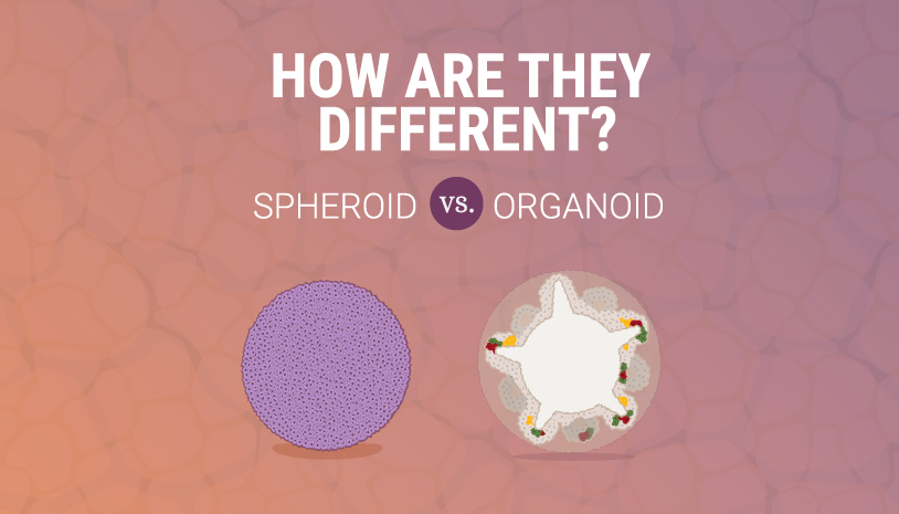
Infographic: Spheroid vs Organoid
Learn the difference between spheroids and organoids in this infographic.
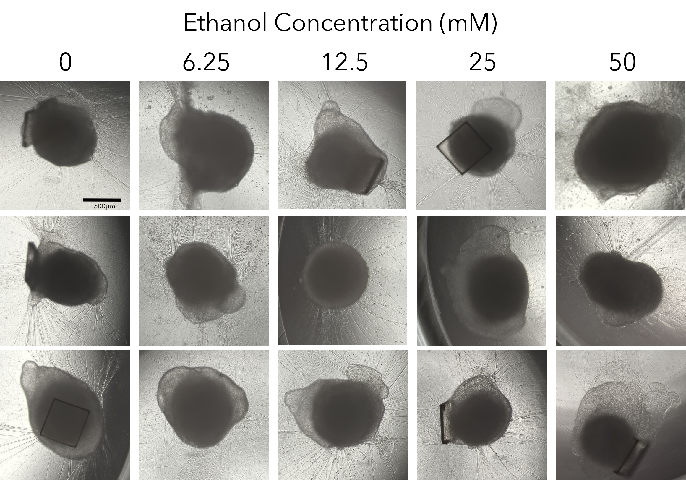
Reproducible Drug Screening Assays Using Single Organoids
Learn how organoid models and assays can be used in high-throughput screening applications.
Why Use 3D Cell Culture?
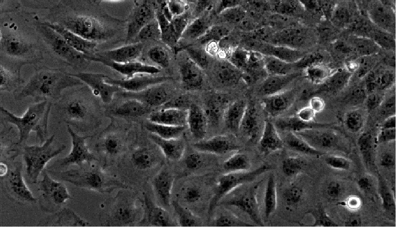
For a long time, scientists have relied on flat 2D cell cultures grown on a plate to study cellular and disease mechanisms. 2D cell models are simple and cost-effective to culture and process. Within the last decade, however, 3D cell cultures have become increasing popular because they are more physiologically relevant and better represent in vivo tissue.
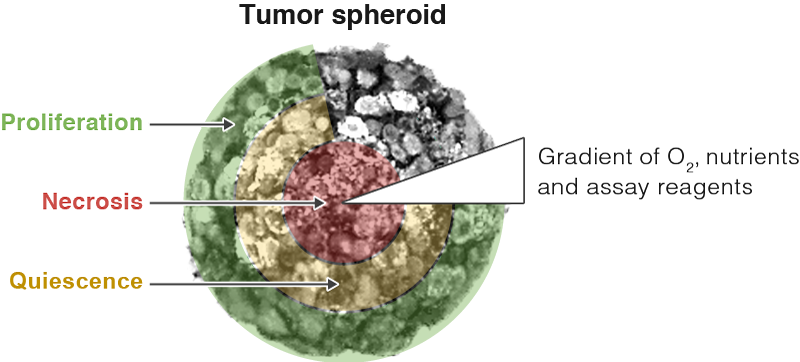
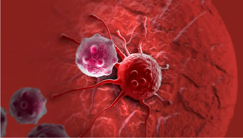
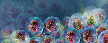
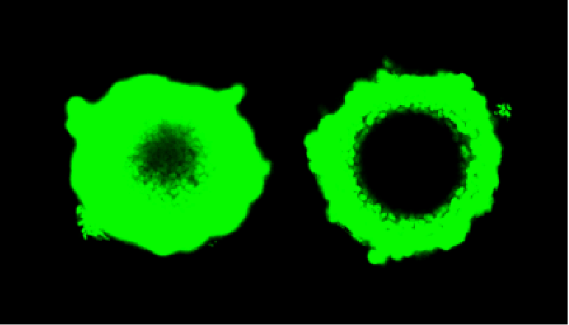
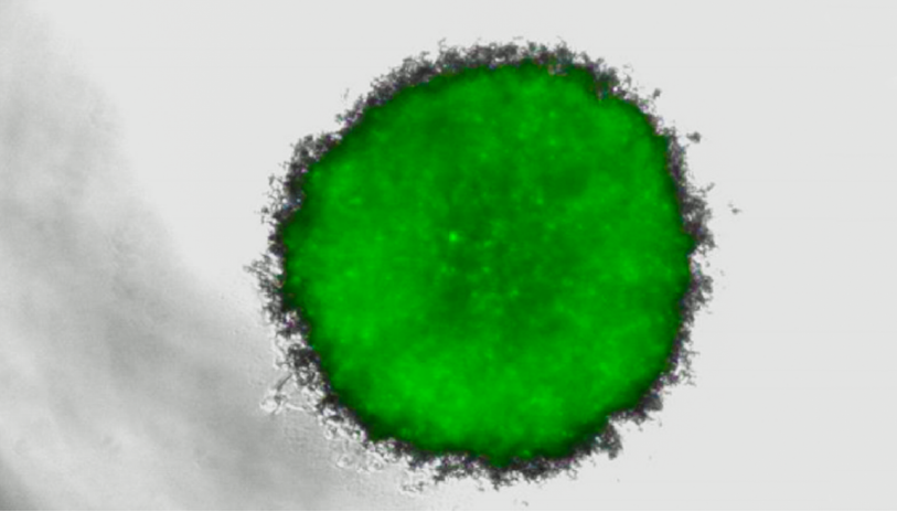
If you think about it, no cell types within our body grow as a monolayer independent of other cells or tissue. Instead, most cells naturally exist in complex 3D structures including different cell types within an extracellular matrix. The numerous cell-cell and cell-matrix interactions all have a profound effect on their behavior. In addition, 2D monolayers have uniform access to nutrients and oxygen, which is not the case in cell masses, such as tumors. 3D tumor spheroids are much more representative of in vivo tumors in which inner cells have less access to nutrients and oxygen compared to the outer layer, forming a natural gradient.
Here are some reasons to use 3D cell cultures:
- 3D cultures better mimic tissue-like structures
- Able to exhibit differentiated cellular function
- Possible to co-culture two or more different cell types
- Can simulate microenvironment conditions such as hypoxia and nutrient gradients
- Better predict in vivo responses to drug treatment
Feature Story: The Promises and Challenges of 3D Models
"That’s what we’re always doing with models: Trying to get them closer to being representative. I think everyone recognizes that 3D is a step forward."
In this feature article, you'll learn about the advantages and challenges of using 3D cell cultures.
3D Cell Culture Applications
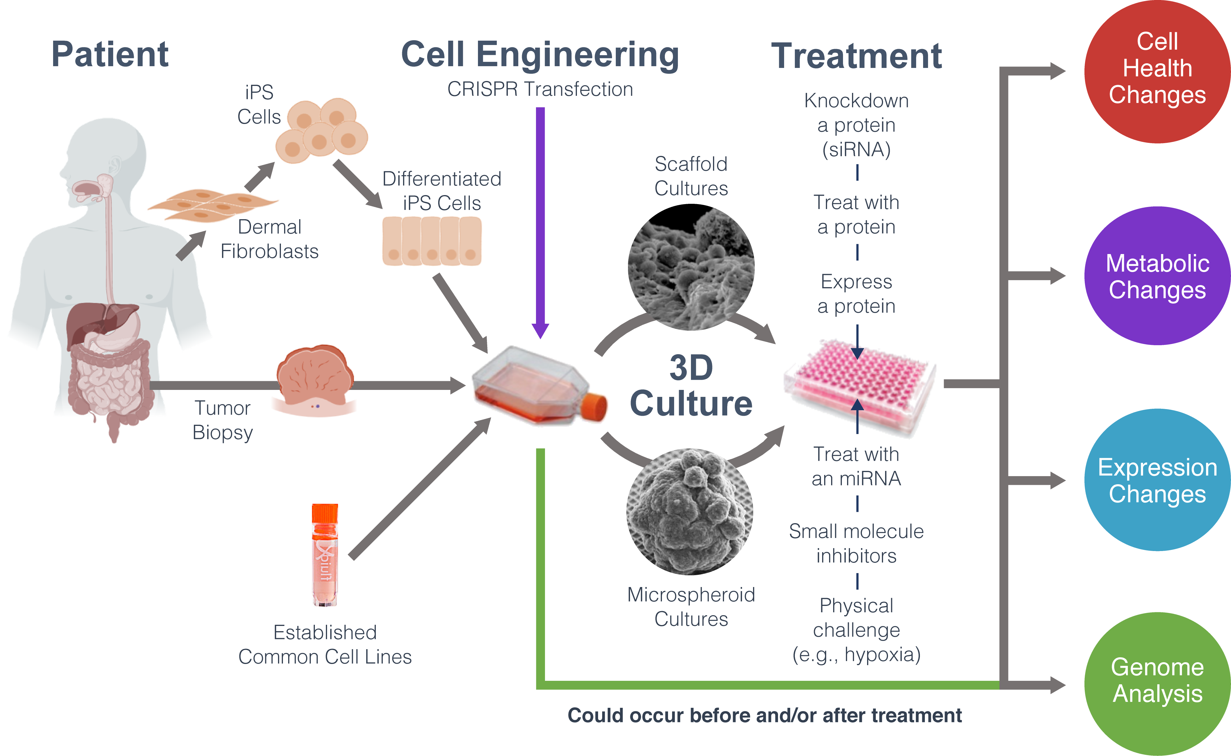
3D cell models are increasingly being used to understand disease mechanisms and discover drug therapeutics. The process may involve deriving 3D cultures, such as cancer organoids, from patients. The 3D cultures can be used to screen for small molecule drugs or genetically manipulated to understand disease pathways. Compared to 2D cultures, 3D cell cultures more accurately predict the efficacy or toxicity of drug treatment.
Monitoring Biology in 3D Cultures
When working with 3D culture models, choosing the right assay system is crucial. Learn about tools for monitoring biology in 3D culture.
How 3D Cultures Are Used in Drug Development
Videos
"I look at developing 3D liver models for nanomaterial assessment...We are aiming to build a battery of models that can reduce or replace animal testing."
"We are developing screens using organoids. We believe they are the next generation 3D model...and offer better predictive outcome in the clinic."
Blog Articles
Evaluating Immunotherapy in Organoids
Read this blog about how patient-derived colorectal organoids can be used to evaluate CAR NK immunotherapy.
Producing Snake Venom with 3D Organoids
Learn how organoids cultured from snake venom glands can be used to develop antivenom.
High-Throughput Drug Screening
Read this blog on how a 3D spheroid-based screening assay is used to find drugs against difficult target proteins.
3D Cell Culture Assays
A big challenge in working with 3D cultures is finding assays that work with your cell type. Most existing cell-based assays were originally designed for 2D monolayers or cells in suspension. Their design may not take into account the size or mass of 3D structures, which may limit cell lysis or overwhelm assay chemistry. In many cases, the extracellular matrix (ECM) coating could prevent reagents from penetrating to the center of the spheroid. This is why it is crucial to optimize and validate cell-based assays for your specific 3D model system. The first steps may include increasing shaking time or incubation time with lysis or detection solutions. Or you may want to choose commercially-available assays that have been qualified to work with a variety of 3D model systems.
Research Highlight: High-Throughput Toxicity Testing in Organotypic Cultures
Learn how high-throughput assays were used to monitor toxicity over time in 3D cell models.
Data: 3D Cell Viability Assays
See how CellTiter-Glo® 3D Assay compares to other viability assays.
Article: Verifying Cell-Based Assays for Use with 3D Models
Assays originally intended for use with monolayer cell cultures need to be adapted to more complex 3D systems.
Story: Determining Cytotoxicity in 3D Liver Spheroids
Learn how the LDH-Glo™ Assay can help predict drug-induced liver injury in 3D microtissues.
Poster: Cell Viability and Cytotoxicity Assays for Tumor Organoids Evaluation
Learn how a variety of bioluminescent and fluorescent assays use different metabolic parameters to assess the viability of tumor organoids.
Webinar: Designing 3D Assays
Learn about factors to consider when designing and optimizing assays for 3D cultures.
Webinar: Method Validation and 3D Islet Models for Diabetes Research
Learn more about islet microtissues and their benefits for diabetes research.
Webinar: 3D Cell Culture and Assays Q&A
Our experts answer common questions on evaluating 3D assays for use with 3D cultures.
Case Study: 3D Assays for Ovarian Cancer Spheroids
Learn how a Yale researcher is using 3D cell viability assays to discover drugs and targeting methods for more efficient chemotherapy.