Lumit® C1q Binding Assay
Evaluate C1q-antibody binding to assess the initiation of the complement-dependent cytotoxicity pathway
- Improved Immunoassay Format: ELISA alternative
- Saves Time: Reduce hands-on steps with a simple, no-wash protocol
- Better Data: No immobilization to plates, beads or other surfaces required
- Results in as little as 60 minutes
Catalog Number:
Size
Catalog Number: CS3695A04
The Ideal Assay to Complement Your Antibody Development Efforts
The Fc region of monoclonal antibodies mediate several effector functions, including complement-dependent cytotoxicity (CDC), through activation of the classical complement pathway. C1q binding to the Fc region is the critical first step in activating this pathway. By quantifying Fc-C1q binding, antibody developers can precisely profile C1q-antibody interactions, predict complement activation kinetics, and accelerate therapeutic antibody selection to ensure both efficacy and safety in therapeutic applications.
Traditional methods to measure C1q binding face several limitations:
- Time-consuming incubation steps
- Highly variable fluorescent readouts
- Low throughput assay set-ups
What does using the Lumit® C1q Binding Assay mean for you?
- Get simple, reliable surrogate measurement of CDC potential in under two hours
- Analyze multiple antibody isotypes and fine-tune your Fc engineering efforts and select promising candidates quickly
- Balance efficacy with safety: achieve sufficient complement activation while minimizing off-target toxicity
- Run your samples in a high-throughput and automation friendly design
How Does the Assay Work?

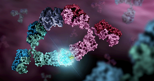
Principle of the Lumit® C1q Binding Assay: The assay provides a quantitative readout of C1q binding to antibody Fc as a proxy for classical complement engagement potential. In a homogeneous, solution-phase format, a biotinylated anti-human IgG Fab is pre-complexed with streptavidin–SmBiT to multivalently capture IgG and induce Fc clustering. C1q–LgBiT is then added; when C1q binds the clustered Fc domains, LgBiT and SmBiT are brought into proximity to reconstitute active NanoBiT® luciferase, producing a luminescent signal proportional to the amount of C1q-bound antibody.
What Is the Assay Workflow?

Lumit® C1q Binding Assay design:
- Biotinylated anti-human IgG Fab is pre-complexed with genetically fused Streptavidin-SmBiT, which multivalently captures IgG in solution and induces Fc clustering.
- Upon C1q-LgBiT addition and IgG binding, SmBiT and LgBiT are brought together, reconstituting active NanoBiT® luciferase.
- The luminescent signal is proportional to C1q bound to the antibody, enabling quantification of antibody-C1q interactions.
Accurately Detect Binding Differences Across Antibody Variants

Lumit® C1q binding assay is suited for distinguishing antibody specificity and sensitivity. The Lumit® C1q Binding Assay was tested against a panel of Rituximab variants that included different IgG isotypes as well as fucoslyation and glycosylation modifications. The binding profiles align with published literature.
Compare for yourself: Impact of structural modifications of IgG antibodies on effector functions.
| Rituximab | Non-fucosylated Rituximab | Non-Glycosylated Rituximab | Rituximab IgG2 isotype | Rituximab IgG3 isotype | Rituximab IgG4 isotype | |
| EC50 (nM) | 9.192 | 8.790 | 5278040 | 57.47 | 2.455 | 38534 |
Interested in other Fc effector functions like ADCC or ADCP?
We offer a wide range of Lumit® binding assays to measure interactions between Fc receptors and antibodies.Protocols
No protocols available
Specifications
Catalog Number:
What's in the box?
| Item | Part # | Size |
|---|---|---|
Human C1q-LgBiT |
CS3695A01 | 1 × 60μl |
Streptavidin-SmBiT |
CS3695A02 | 1 × 30μl |
Anti-human IgG Fab-Biotin |
CS3695A03 | 1 × 150μl |
Lumit® Immunoassay Dilution Buffer A, 10X |
VB201A | 1 × 10ml |
Lumit® Detection Substrate A |
VB301D | 1 × 75μl |
Resources
Featured Resource: Webinar
Lumit® Immunoassays: An Easier Faster Method for Analyte Detection
In Lumit® Immunoassays, antibodies (or other affinity reagents) are chemically labeled with the small and large subunits of NanoLuc® Luciferase, known as SmBiT and LgBiT, respectively. In the presence of an analyte, the two antibodies come into close proximity, allowing SmBiT and LgBiT to form an active enzyme and generate a bright luminescence signal.
- Use simple, add-mix-read protocols to detect a variety of analytes
- Implement existing Lumit® assays in your lab or build your own Lumit® immunoassays
- Analyze data using examples for cytokine detection, signaling pathway analysis and FcRn binding
Related Products
Similar Products
Lumit® Immunoassay Cellular Systems
A no-wash bioluminescent immunoassay to measure levels of target analytes in cell lysates.
W1201, W1202, W1203, W1331, W1332, W1333, W1220
Lumit® Immunoassays for Detection of Metabolic Regulators
Assays to detect insulin or glucagon in cell culture samples with a scalable, no-wash protocol.
Frequently Used With
Lumit® FcγR Binding Immunoassays
Novel homogeneous competition assays to measure the interaction between human Fc receptors and antibodies or Fc fusion proteins.
W7030, W7031, W7040, W7041, W7050, W7051, W7060, W7061, W7070, W7071, W7080, W7081
Lumit® FcRn Binding Immunoassay
A novel homogeneous (no-wash) competition assay to measure the interaction between human FcRn and Fc proteins, including antibodies.
W1151, W1152
ADCC Reporter Bioassay, V Variant
Measure potency and stability of antibodies mediating ADCC through the high-affinity human FcγRIIIa-V receptor.
G7015, G7014, G7010, G7016, G7013, G7018, G7102, GA1130
ADCP Reporter Bioassay (THP-1)
Measure the potency and stability of antibodies and other biologics with Fc domains that bind and activate FcγRs.
JA9411, JA9415, GA1272, GA6020