Lumit® Immunoassays for Research
Lumit® Immunoassays provide a simple and fast alternative to conventional immunoassay methods including sandwich ELISAs and Western blots. Lumit® assays are sensitive, have broad dynamic range and can be completed in as little as 30 minutes, making them a compelling alternative to labor-intensive ELISA and Western blot methods.
Advantages of Lumit® Assays over conventional immunoassays include:
- Simple add-mix-read protocol with no washing steps
- Direct analyte measurement in the cell culture plate or on medium removed from the cells
- No immobilization to plates, beads or other surfaces required
- Sensitive luminescence detection with wide dynamic range
Lumit® Assays are available as catalog items, early-access materials or by custom order.
How Lumit® Immunoassays Work
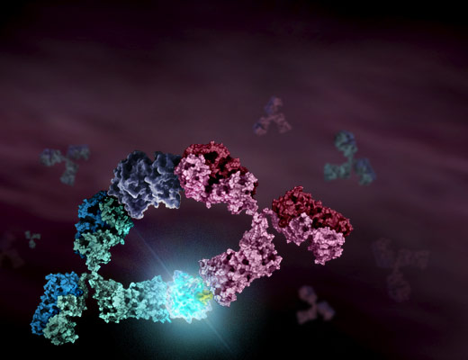
Lumit® Immunoassays are based on NanoLuc® Binary Technology (NanoBiT®). In Lumit® Immunoassays, antibodies are chemically labeled with the small and large subunits of NanoLuc® Luciferase, known as SmBiT and LgBiT, respectively. In the presence of an analyte, the two antibodies come into close proximity, allowing SmBiT and LgBiT to form an active enzyme and generate a bright luminescence signal.


Filter By
Shop Lumit® Immunoassay Products
Showing 25 of 25 Products
Lumit® Immunoassays Available by Custom Order
In addition to the early-access materials listed above, we offer consultation on development of customized Lumit® Assays. Please contact us to discuss at the link below to discuss development of a custom solution.
What are Lumit® Immunoassays?
Lumit® Immunoassays are based on the chemical labeling of antibodies and proteins with SmBiT and LgBiT using HaloTag® technology. In the presence of an analyte, the two antibodies come into close proximity and allow SmBiT and LgBiT to form an active enzyme and generate a bright luminescence signal proportional to analyte concentration. They provide a fast alternative to traditional immunoassays, such as ELISA and Western blotting.
The Lumit® Immunoassay Labeling Kit is used to label pairs of antibodies to create Lumit® Assays. Lumit® Detection Reagents and prelabeled LgBiT and SmBiT secondary antibody conjugates are also available.
Lumit® Immunoassays for cytokine detection quantitatively measure target analytes in cell culture samples with a simple, no-wash assay protocol. The assays can be performed directly on cell culture samples or on medium transferred from cell plates.
In the Lumit® Immunoassay Cellular System, the LgBiT and SmBiT subunits are conjugated to a pair of secondary antibodies from two different species. In a multiwell plate, seeded cells are lysed using a NanoBiT-compatible lysis buffer (digitonin), and the target protein is detected by adding an antibody mix containing two primary antibodies against the protein along with the SmBiT- and LgBiT-conjugated secondary antibodies.
The Lumit® FcRn Binding Immunoassay is a competition assay to measure the interaction between human FcRn and Fc proteins, including antibodies. A Human IgG1 labeled with LgBiT (Tracer-LgBiT) is used as the tracer. A C-terminal biotinylated human FcRn bound to Streptavidin-SmBiT is used as the target. In the absence of an antibody analyte sample, Tracer-LgBiT binds to the hFcRn-SmBiT target resulting in maximum luminescence signal. Unlabeled IgG will compete with Tracer-LgBiT for binding to the FcRn target, resulting in a concentration dependent decrease in luminescent signal.


