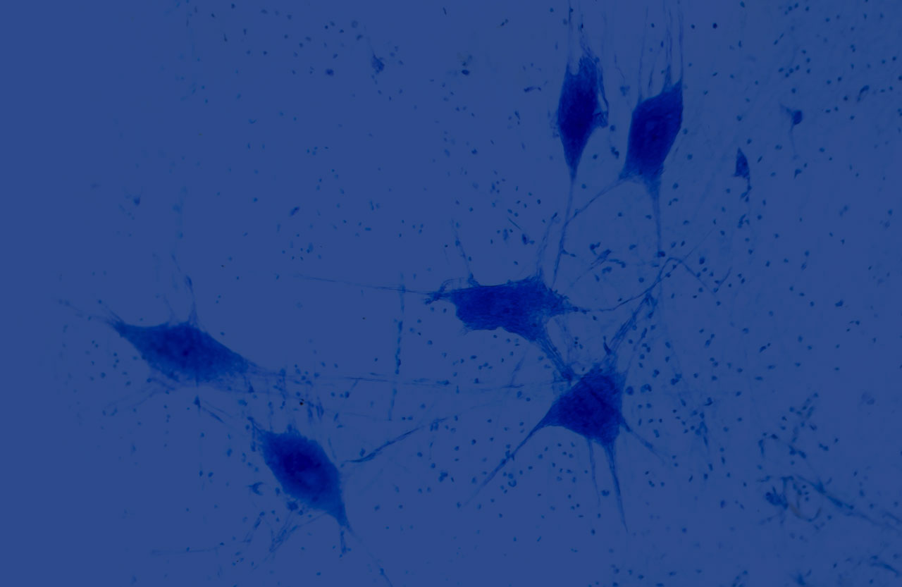
Neuroscience Research Solutions
The field of neuroscience seeks to understand how billions of interconnected neurons give rise to memory, behavior, and human health.
We support your journey by providing a comprehensive portfolio of bioluminescent and fluorescence-based assays, genetic reporters, and streamlined workflows. These tools are designed to reveal what’s happening inside the cell without slowing your pace.
Together, we can decode the brain’s complexity—transforming quantitative insights into meaningful scientific progress.
Tools for Neural Organoids
Organoids offer a physiologically relevant, three-dimensional model for studying the human brain. They provide a powerful platform for studying neurodevelopmental and neurodegenerative disorders, as well as evaluating drug responses in a controlled yet complex environment.
We provide assay technologies optimized for the demands of organoid-based neuroscience:
- Real-time, non-invasive health monitoring using bioluminescent viability and cytotoxicity assays
- Functional analysis via live-cell biosensors that measure key signaling events and neuronal activity
- High-sensitivity detection platforms for biomarker discovery and drug screening in low-volume, complex samples
Measure Neurotoxicity with Sensitive and Scalable Assays
Assessing cell viability and cytotoxicity is essential for evaluating neuronal health, particularly in complex or sensitive models such as neural organoids and stem cell–derived neurons.
Our luminescent assays can measure:
- Drug effects and potential neurotoxicity
- Metabolic activity shifts (e.g., ATP levels)
- Membrane integrity loss (e.g., LDH release)
- Long-term culture performance and stability
The advantages of luminescent assays include:
- Non-destructive formats that preserve sample integrity
- High sensitivity and reproducibility, even in low-density cultures
- Compatibility with high-throughput and longitudinal workflows
Featured Publication
Learn how the LDH-Glo Cytotoxicity Assay can reliably assess viability and cytotoxicity in cerebral organoids regardless of organoid size or cell density in this publication: Development and optimization of a lactate dehydrogenase assay adapted to 3D cell cultures.
Track Proteins in Live Neurons
In neuroscience research, precise and minimally invasive tools for tracking protein behavior in live cells are essential for understanding complex processes such as synaptic plasticity, protein aggregation, and neuronal differentiation.
Our HiBiT and HaloTag technologies offer complementary approaches for tagging and analyzing endogenous proteins in neurons and neural organoids. The table below compares their features and applications to help guide your selection based on experimental needs.
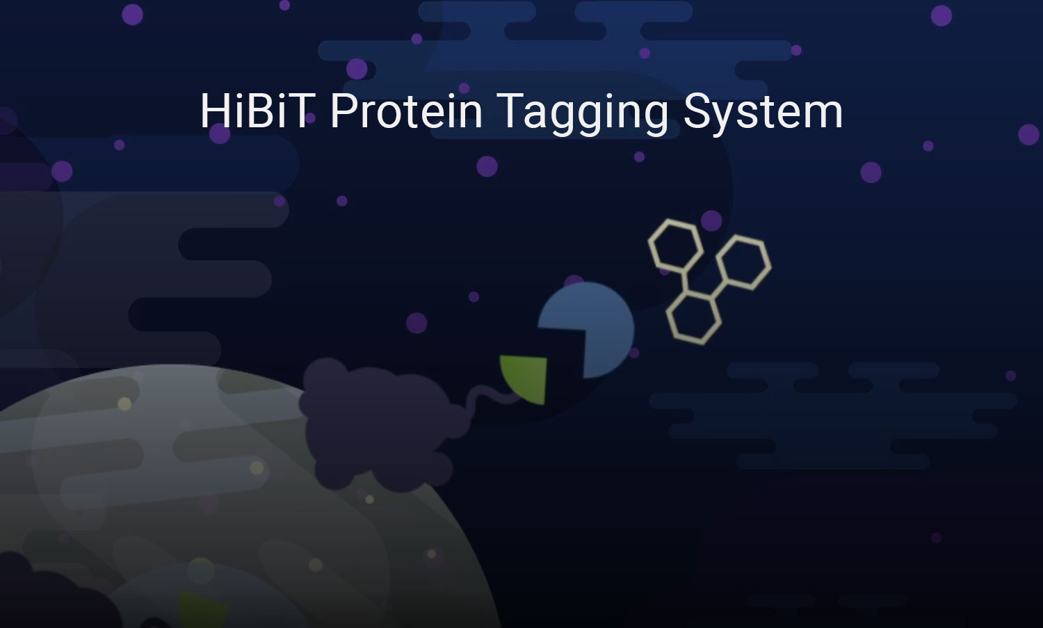
What is HiBiT?
What is HaloTag?
| Tag | HiBiT | HaloTag |
|---|---|---|
| Detection Method |
Luminescence (no excitation light required) |
Fluorescence (requires external illumination) |
| Key Applications |
|
|
| Labeling Mechanism | Reconstitution of luciferase with complementary LgBiT fragment | Covalent binding to synthetic ligands (e.g. fluorophores, biosensors) |
| Tag Size | 11 amino acids | 297 amino acids |
| Live-cell Compatibility | Yes. Supports real-time, non-invasive measurements in live neurons and organoids | Yes. Enables high-resolution imaging in live cells and organoids |
| Phototoxicity | Low. No light excitation needed | Potential for phototoxicity depending on light exposure |
| Integration with CRISPR | Yes. Ideal for tagging endogenous proteins with minimal footprint | Yes. Allows precise targeting of ion channels and signaling proteins |
| Single-molecule Imaging | Not ideal | Well-suited. Supports super-resolution and single-molecule imaging |
Neural Cell Imaging
Imaging plays a foundational role in neuroscience, enabling researchers to explore everything from intracellular signaling to complex neural circuits. High-resolution, live-cell imaging is especially valuable to visualize synaptic activity, track neuronal signaling, and explore disease mechanisms in real time. However, traditional imaging techniques often come with limitations, including:
- Phototoxicity that can damage sensitive cells during observation
- Limited sensitivity, which restricts detection of low-abundance signals
- Cumbersome workflows, which can hinder reproducibility and scalability
To overcome these challenges, we offer novel fluorescent ligands and bioluminescence imaging tools that empower you to explore the brain with greater clarity, precision, and confidence.
High-Resolution Imaging with Reduced Background and Enhanced Stability
Our Janelia Fluor® HaloTag® Ligands are designed for advanced imaging of HaloTag® fusion proteins in live-cell, fixed, and endogenous environments—making them ideal for neuroscience research.
Spanning the visible spectrum, these dyes feature fluorogenic properties that enhance brightness and reduce background, enabling clear, high-resolution imaging of neuronal structures and processes.
Key benefits for neuroscience workflows:
- Compatible with super-resolution, confocal, in vivo, and live-cell imaging
- High photostability minimizes bleaching—ideal for long-term studies of neurodegeneration and plasticity
- Pulse-chase labeling supports real-time tracking of protein dynamics, including trafficking and turnover
Visualize Low-Abundance Protein Dynamics in Live Cells
Dynamic cellular processes—such as protein localization, signaling, and cell-to-cell variability—play critical roles in brain development, function, and disease. Our real-time imaging tools including luciferase reporters and bioluminescent imaging system can help uncover these complex mechanisms in neural systems.
Luciferase Reporter
NanoLuc® Luciferase is a small, bright, and highly stable bioluminescent enzyme that is ideal for studying complex, dynamic biological processes in live cells.
Advantages to using NanoLuc for neuroscience research include:
- High sensitivity enables detection of low-abundance neural signals and proteins
- No excitation light eliminates phototoxicity—ideal for sensitive neurons and long-term assays
- Real-time, kinetic readouts for tracking signaling, gene expression, and protein interactions
- Compatible with modular systems like NanoBiT and HiBiT for studying protein dynamics
- Low background and scalable format support reproducible, high-throughput neuroscience workflows
Bioluminescence Imaging System
The GloMax® Galaxy Bioluminescence Imager System enables real-time imaging of NanoLuc® Luciferase signals to monitor protein dynamics and cellular physiology. It is compatible with NanoLuc® workflows, allowing you to visualize cell-to-cell differences within neural populations and track intracellular dynamics.
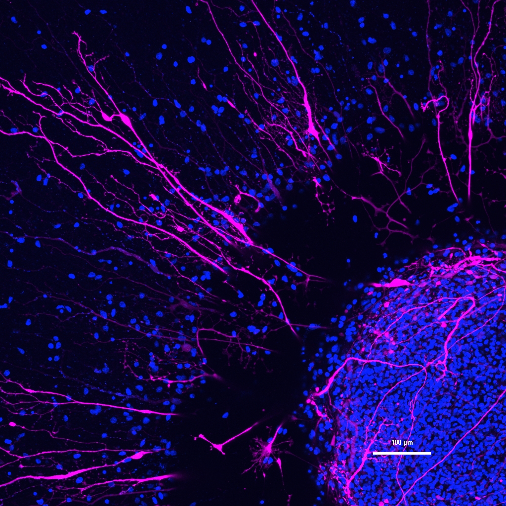
See the Data: Visualizing GPCR Target Engagement

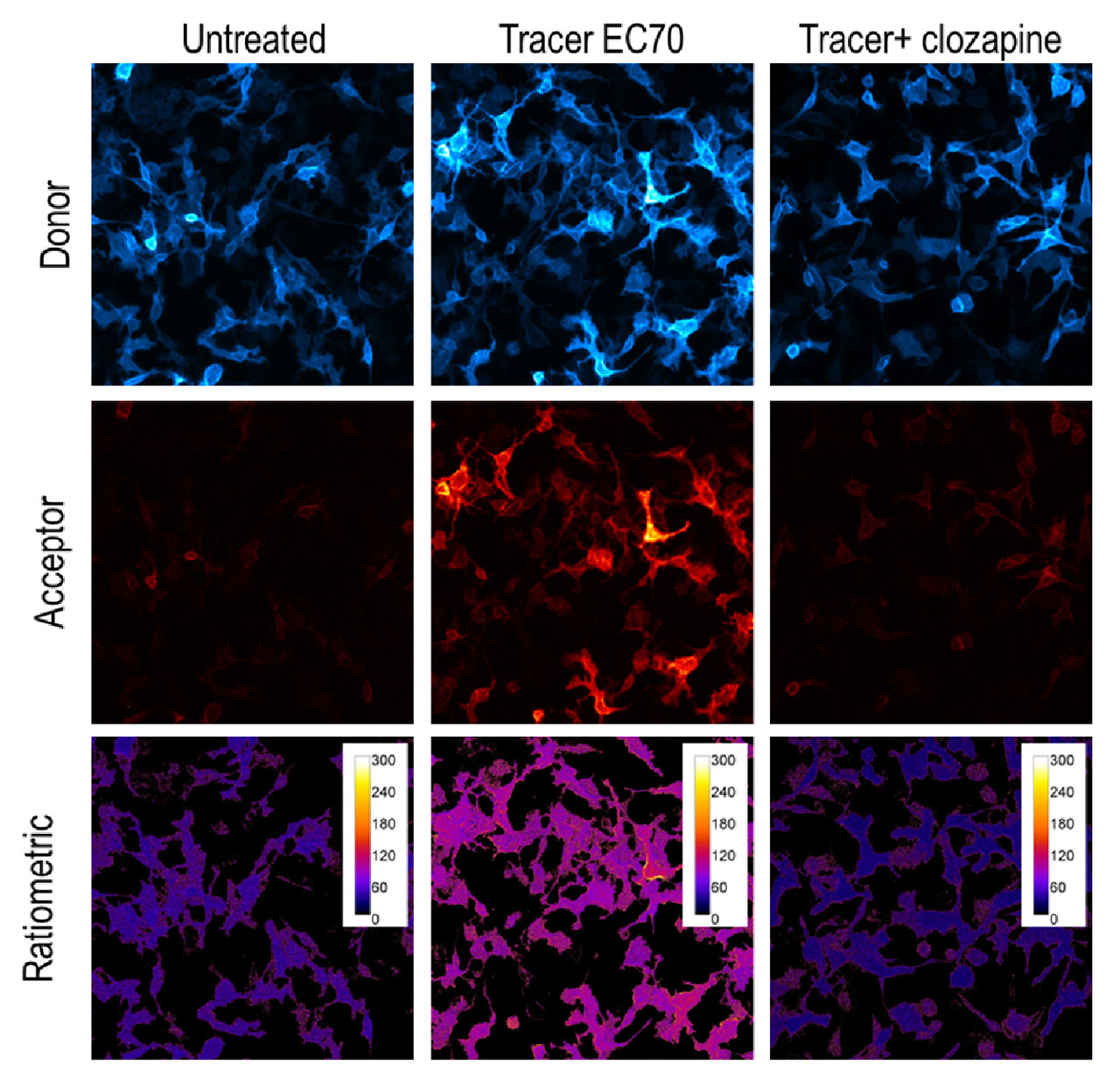
GPCR Target Engagement for HTR2C Receptor using NanoBRET. Left Panel: HiBiT-5-HR2C target displacement using either clozapine or serotonin measured on the GloMax® Discover Microplate Reader. Right Panel: Donor and acceptor images captured on the GloMax® Galaxy Bioluminescence Imager. The donor channel was exposed for 60 seconds and acceptor channel was exposed for 120 seconds. Ratiometric images demonstrate BRET-ratio upon addition of Tracer EC70 alone or in combination with clozapine.
Image the Brain in Live Animals
Due to its high brightness and low background, NanoLuc is ideal for non-invasive imaging in whole organisms, including the brain.
Nano-Glo® Cephalofurimazine (CFz9) In Vivo Substrate is designed to cross the blood-brain barrier, enabling the detection of NanoLuc® Luciferase in the brain of living animals. It supports applications such as:
- In vivo monitoring of gene expression or promoter activity in specific brain regions
- Longitudinal studies of neurodegenerative disease progression
- Studies of therapeutic distribution to the brain
- Chronic monitoring of neural circuits in animal models
See more resources on neural cell imaging:
Webinar: Real-Time Non-Destructive Monitoring of Human Cerebral Organoids
Learn how the NanoLuc®-HaloTag® Reporter System and Janelia Fluor® HaloTag® Ligands enable continuous monitoring of organoid formation and neural differentiation.
Poster: Real-Time Analysis of Psychedelic Activity in Neural Models
Learn how the NanoLuc®-HaloTag® Reporter System is used to monitor effects of psychedelic tryptamines on human neural stem cells.
Article: Imaging Advanced Cell Models
Learn how HaloTag® technology enables long-term, high-resolution imaging of protein dynamics in complex 3D neural models like cerebral spheroids.