Illuminating Transmembrane Proteins Using HiBiT CRISPR Cell Lines
Promega Corporation
Publication date: May 2025
Introduction
HiBiT technology is a powerful tool for studying transmembrane and extracellular proteins, featuring a small, luminescent tag suitable for sensitive and quantitative detection of protein expression, localization, and dynamics. HiBiT is an 11-amino-acid fragment of the NanoBiT® complex, which, upon binding to its complementary partner LgBiT, reconstitutes a functional luciferase enzyme. Addition of substrate generates a luminescent signal strong enough to detect proteins at their endogenous levels. The minimal size of HiBiT reduces interference with the native function of tagged proteins and facilitates precise CRISPR-based tagging, thus avoiding artifacts common in overexpression models. Additionally, NanoBiT® luciferase activity is compatible with both intra- and extracellular environments, making it particularly effective for analyzing transmembrane proteins. In this article, we demonstrate the utility of HiBiT by tracking Epidermal Growth Factor Receptor (EGFR), a transmembrane receptor tyrosine kinase essential for cell proliferation signaling.
Generating a HiBiT-EGFR CRISPR Clonal Cell Line
We generated endogenous HiBiT-tagged EGFR cells via CRISPR knock-in. Tagging genes at their endogenous locus ensures proteins are expressed at native levels under native regulatory control which is crucial for accurately studying biologically relevant processes such as receptor signaling and trafficking. We selected a knock-in site between the native signal peptide and the N-terminus of the mature EGFR protein (residues 24/25) to preserve proper trafficking and HiBiT integrity up signal peptide removal. When the native signal peptide is unknown or undefined, as is the case for approximately 80% of GPCRs, an alternative, well-characterized signal peptide (e.g., IL-6) can be introduced alongside HiBiT. However, ensuring that only a single signal peptide is present is essential for correct protein trafficking.
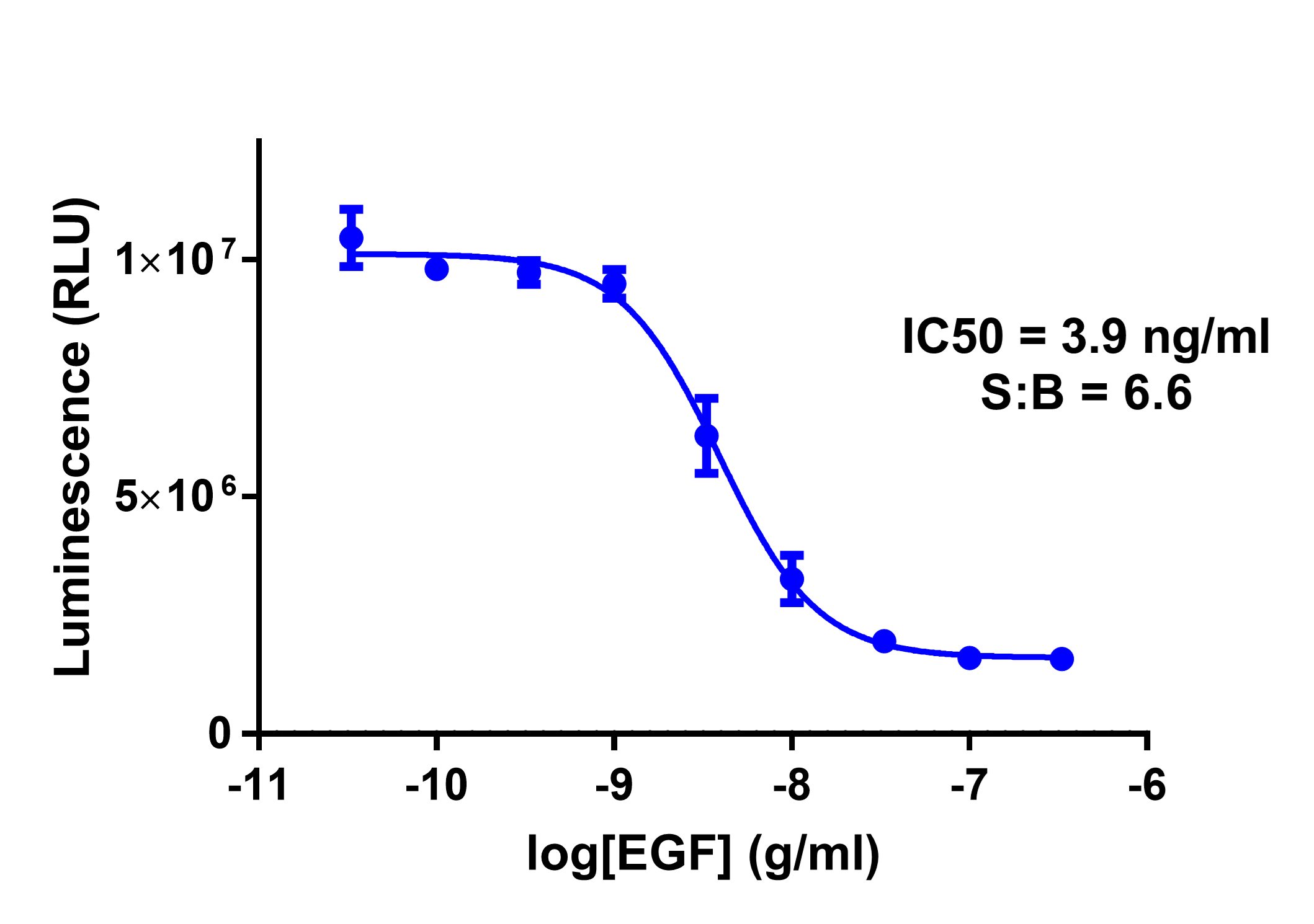
When HiBiT is presented extracellularly, live-cell flow cytometry using Anti-HiBiT Monoclonal Antibody, XFD Fluorophore Conjugates, enables enrichment of CRISPR-edited pools for HiBiT-positive cells. Here, flow cytometry results revealed a cell pool contained approximately 10% HiBiT-EGFR cells (Figure 1, red trace). A single HiBiT-EGFR clonal cell line was then established following sequencing and allelic counting to verify no additional mutations occurred during the gene editing. Subsequent flow cytometry confirmed robust surface HiBiT-EGFR expression in the clonal line (Figure 1, orange trace), validating that HiBiT tagging did not disrupt EGFR trafficking to the plasma membrane.

Quantification of EGFR Internalization
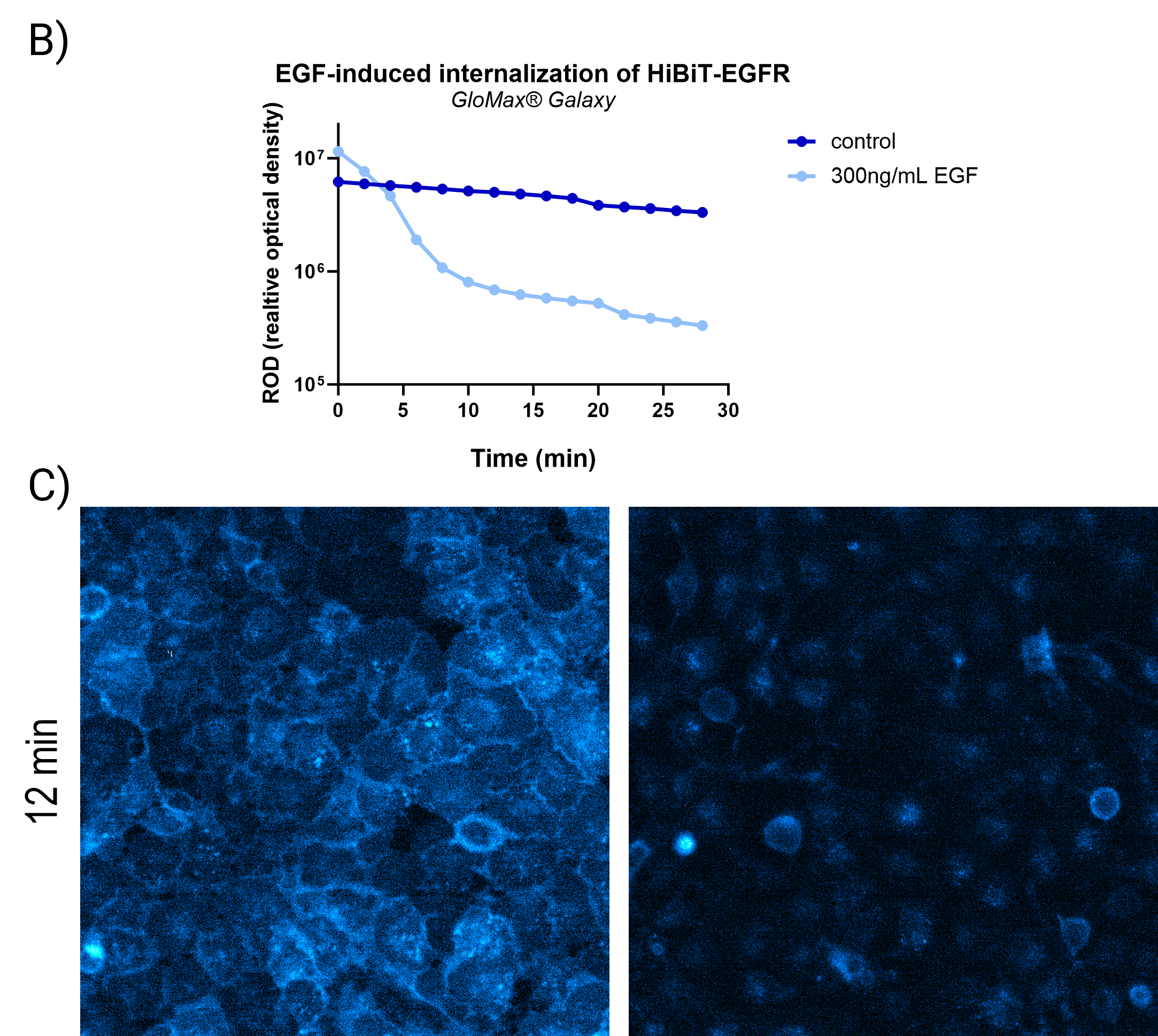
Internalization is a critical mechanism regulating the signaling and degradation of EGFR. After ligand binding, EGFR undergoes endocytosis, followed by recycling or lysosomal degradation, processes that precisely control signaling intensity and duration. Disruption of EGFR internalization can lead to prolonged signaling, contributing to uncontrolled cellular growth and survival commonly observed in cancers (1,2).
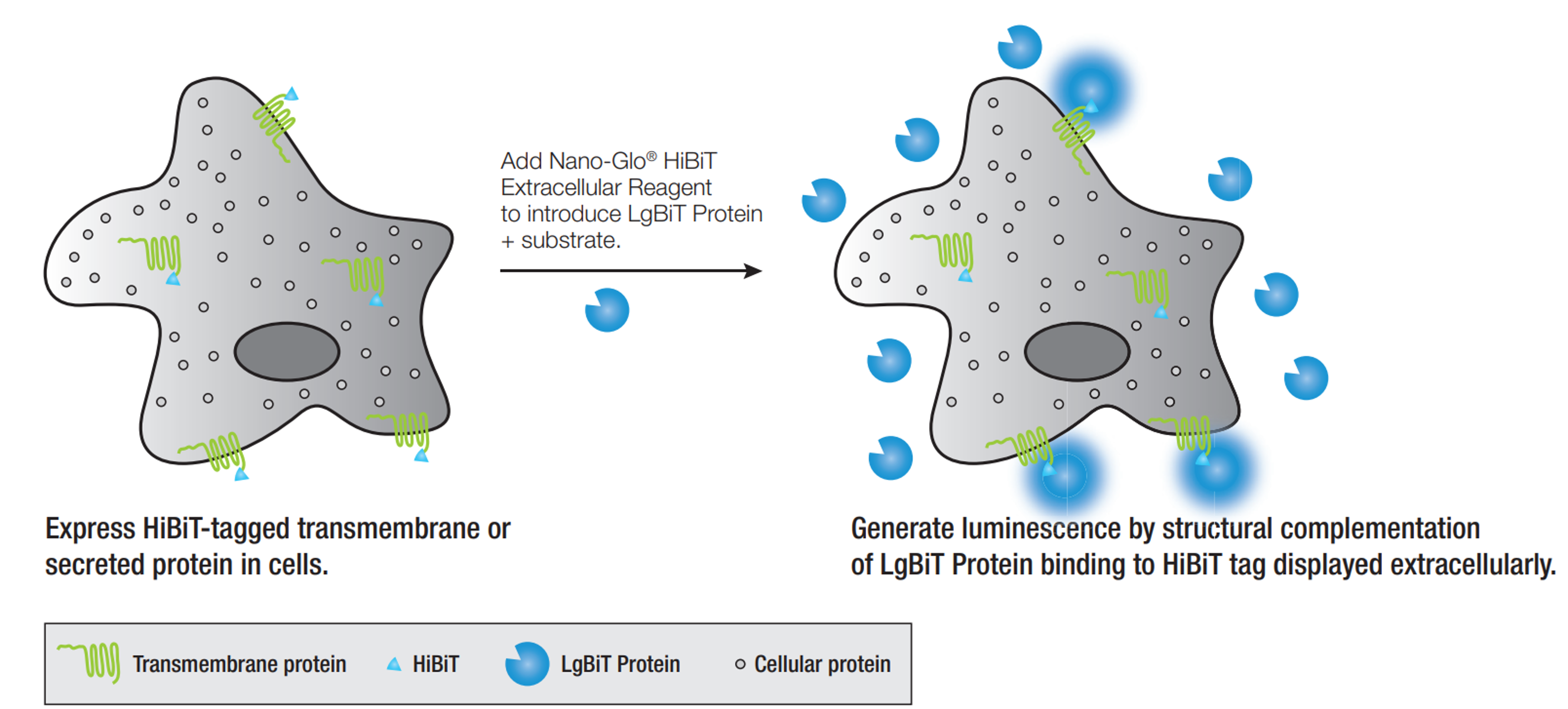
Nano-Glo® HiBiT Extracellular Detection System measures surface expression of HiBiT-tagged EGFR by introducing substrate and purified, cell-impermeable LgBiT directly into live-cell culture media (Figure 2). LgBiT selectively binds to extracellular HiBiT, producing a luminescent signal specific to EGFR localized on the plasma membrane. Internalization removes HiBiT-EGFR from the extracellular environment, decreasing luminescence proportionally to the amount internalized. Nano-Glo® HiBiT Extracellular Detection System facilitates rapid and quantitative assays, either in a plate-based format or through imaging using the GloMax® Galaxy Bioluminescence Imager (For Research Use Only).
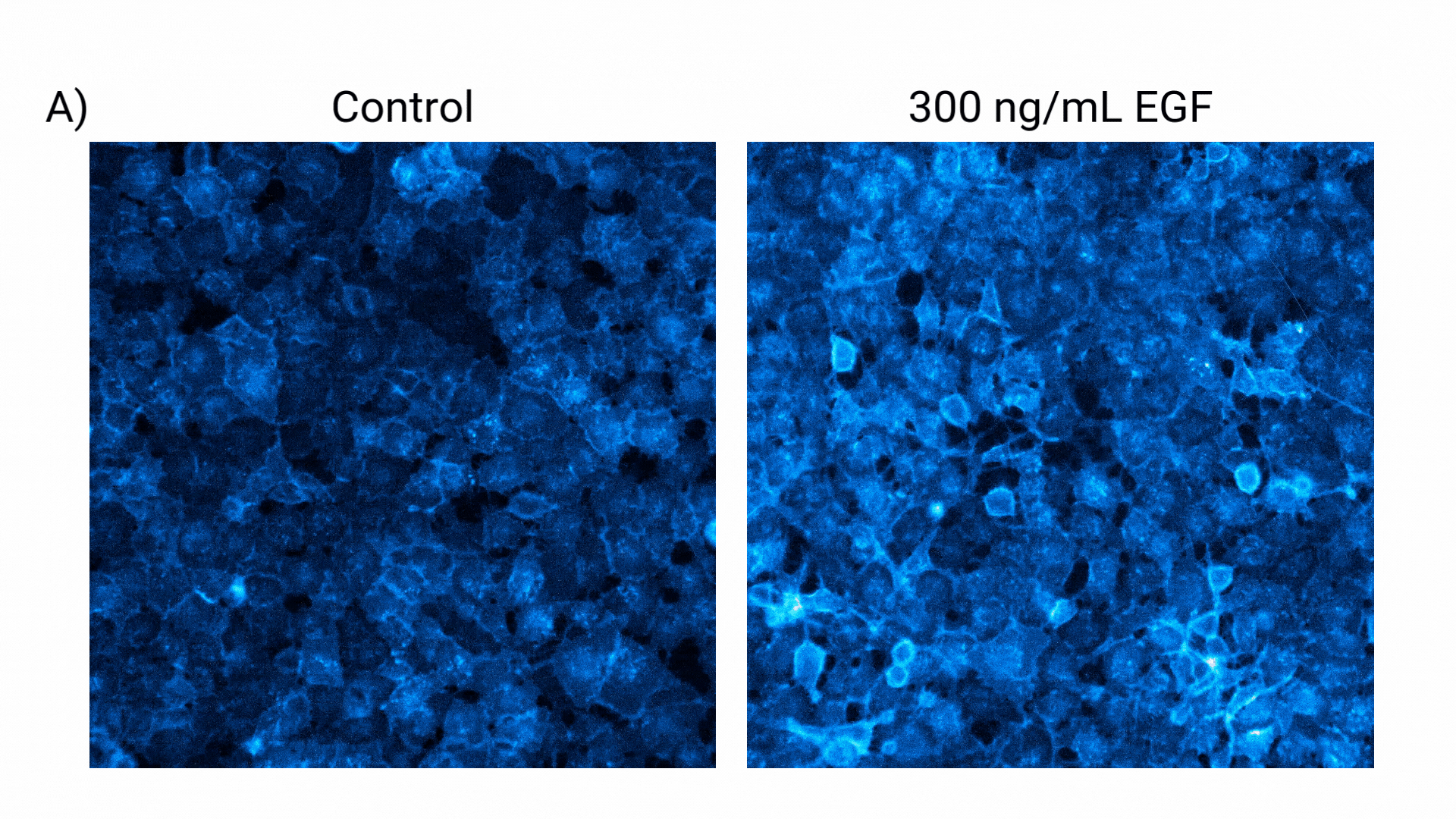
Visualization of EGFR Internalization
Conclusions
These methods offer powerful, real-time, non-invasive approaches to monitor receptor dynamics at the cell surface, providing insights into receptor trafficking and regulation. The Nano-Glo® HiBiT Extracellular Assay’s high sensitivity, quantitative, and scalable nature make it particularly useful for studying this dynamic process, which plays a critical role in cellular signaling and disease biology.
Citations
- Roepstorff, K. et al. (2008) Endocytic downregulation of ErbB receptors: mechanisms and relevance in cancer. Histochemistry and Cell Biology 129(5), 563–78.
- Sigismund, S. et al. (2018) Emerging functions of the EGFR in cancer. Molecular Oncology 12(1), 3–20.
- Sorkin, A. and Goh, L.K. (2009) Endocytosis and intracellular trafficking of ErbBs. Experimental Cell Research 315(4), 683–96.
- Fortian, A. and Sorkin, A. (2010) Live-cell fluorescence imaging reveals high stoichiometry of EGFR endocytosis. Journal of Cell Science 123(Pt 19), 3213–21.
Curious about HiBiT Protein Tagging?
Interested in CRISPR Knock-In Tagging?
Related Resources

Technical Article: Quantifying Percent Surface Expression using Bioluminescent Detection
Learn how to use the HiBiT tag to quantify the percent surface expression of a protein.
Technical Article: HiBiT-Based Detection of PCSK9 Secretion
Read about how HiBiT expands protein trafficking studies.