Anti-HiBiT Monoclonal Antibody
Sensitive Antibody-Based Detection of HiBiT-Tagged Proteins
- Use as an orthogonal method to verify bioluminescent results
- Confirm subcellular localization of endogenous HiBiT-tagged proteins in cell pools or clones following CRISPR/Cas9 HiBiT knock-in
- Confirm protein levels and size using classic Western blot detection
- Perform immunoprecipitation of HiBiT-tagged proteins
- Perform FACS analysis on live cells (extracellular HiBiT) or on fixed cells (intracellular HiBiT)


Catalog Number:
Size
Catalog Number: N7200
Catalog Number: N7210
Immunodetection of HiBiT-Tagged Proteins
The 11-amino-acid HiBiT peptide tag can be added to a protein of interest using traditional cloning or CRISPR/Cas9 genome editing, enabling analysis of proteins under endogenous expression conditions. HiBiT-tagged proteins are typically measured using the bioluminescent signal generated by adding Nano-Glo® HiBiT detection reagents, providing for precise measurement of changing protein levels over 7 logs of linear dynamic range. Adding the specific and highly sensitive Anti-HiBiT Monoclonal Antibody expands the detection options for HiBiT-tagged proteins, enabling traditional antibody-based methods for detecting the HiBiT tag and removing the need for tandem-tagging approaches.
Specifically Detect Sub-Picogram Levels of HiBiT via Western Blot
The Anti-HiBiT Monoclonal Antibody is highly sensitive and specific, detecting sub-picogram levels of HiBiT-tagged proteins with minimal background in Western blots.
Sensitivity and specificity of Western blotting with the Anti-HiBiT Monoclonal Antibody. HaloTag®-HiBiT protein was serially diluted into K562 mammalian cell lysate to generate samples of 0.125–4pg of HiBiT-tagged protein. After SDS-PAGE, the gel was transferred to a PVDF membrane, and immunoblotting was performed using 1µg/ml of primary antibody, 0.2µg/ml of HRP-conjugated secondary antibody and ECL Western Blotting Substrate. The image was exposed for 60 minutes, and the brightness scaled 1,200–15,000.

Subcellular Localization Following CRISPR/Cas9 HiBiT Knock-In
Immunofluorescent staining using the Anti-HiBiT Monoclonal Antibody allows characterization of HiBiT knock-in cell pools and clones to confirm that the endogenously expressed HiBiT-tagged protein has the expected subcellular localization.
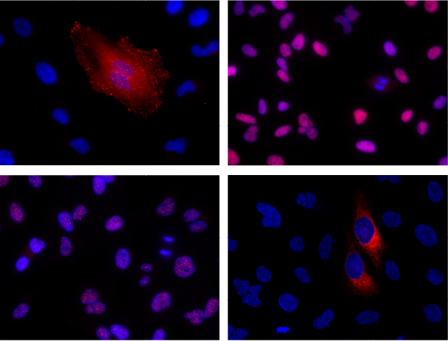
Immunofluorescent detection of HiBiT-tagged proteins in CRISPR-edited cell pools and clones using the Anti-HiBiT Monoclonal Antibody. CRISPR/Cas9 editing was used to knock-in HiBiT at the endogenous locus of proteins with varying subcellular localization. Fixed CRISPR-modified clones or pools of cells were imaged by immunofluorescent staining with the Anti-HiBiT Monoclonal Antibody (red) and Hoechst dye (blue). Panel A. VCL-HiBiT pool. Panel B. HDAC2-HiBiT clone. Panel C. SMARCA4-HiBiT clone. Panel D. HSP90B1-HiBiT pool.
Anti-HiBiT Antibody Performs As Well As or Better Than Common Epitope Tag Antibodies
Western blot detection and immunoprecipitation of HiBiT-tagged proteins demonstrate specific detection and efficient pull-down of HiBiT-tagged proteins with performance similar to or better than FLAG®, Myc or HA tags when using common antibodies.


Comparison of Western blot detection using Anti-HiBiT Monoclonal Antibody to common epitope tag antibodies. HSP90B1 was endogenously tagged on the C-terminus in HeLa cells with HiBiT alone or HiBiT and a single copy of the Myc, HA or FLAG® tags, or three copies of the FLAG® tag. Lysates (10µg) from the HSP90B1-tagged cell pools were separated by SDS-PAGE. After transfer to PVDF membrane, Western blotting was performed with 1µg/ml Anti-HiBiT or Anti-Myc, 0.1µg/ml Anti-HA or 10µg/ml Anti-FLAG® primary antibodies (as indicated) and 0.4µg/ml anti-mouse or anti-rat (for anti-HA blot) secondary antibody and ECL substrate using a 5-minute exposure. The HiBiT antibody image brightness was scaled 0–29,817 and all other images 145–2735.


Comparison of pull-down efficiency from CRISPR-modified cell pools. CRISPR/Cas9 gene editing was used to knock in either HiBiT, HiBiT-FLAG, HiBiT-3XFLAG, HiBiT-HA or HiBiT-Myc to the C-terminus of the endogenous locus of the HSP90B1 protein. Lysates from pools of CRISPR-edited HeLa cells were diluted into buffer and split to generate equivalent samples for pull-down comparisons. The HSP90B1 protein was immunoprecipitated with magnetic Protein G resin (Cytiva) using 2µg of either the Anti-HiBiT, FLAG®, HA or Myc antibody as indicated. After a 2-hour incubation, resins were washed, and the proteins were eluted by heating in SDS loading buffer. The amount of tagged HSP90B1 protein precipitated from cell lysates was detected using the Nano-Glo® HiBiT Blotting System. Both eluted protein and the protein remaining in the supernatant are shown. The intensity of the signal in the starting sample compared to the antibody-treated supernatant indicates the protein clearance efficiency of the antibody.
Protocols
Complete Protocol
Specifications
Catalog Number:
Contenido
| Item | Part # | Presentación |
|---|---|---|
Anti-HiBiT Monoclonal Antibody |
N720A | 1 × 100μg |
SDS
Search for SDSCertificado de Análisis
Use Restrictions
For Research Use Only. Not for Use in Diagnostic Procedures.Condiciones de Almacenaje
NOT FOR MEDICAL DIAGNOSTIC USE. FOR IN VITRO USE ONLY. BY USE OF THIS PRODUCT, RESEARCHER AGREES TO BE BOUND BY THE TERMS OF THIS LIMITED USE LABEL LICENSE. If researcher is not willing to accept the terms of this label license, and the product is unused, Promega will accept return of the unused product and provide researcher with a full refund.
This product and its derivatives may not be further transferred by the researcher and the purchased quantity of the product may be used only by the researcher, and then only for (1) research use, which may include drug discovery and development research; and (2) use in provision of services, that result in transfer of information or data only. No other commercial use of this product or derivatives is allowed. “Commercial use” means any and all uses of this product or derivatives by a party in exchange for money or other consideration, including, but not limited to, (1) use in further product manufacture; and (2) resale of the product or its derivatives, whether or not such product or derivatives are resold for use in research. Researcher may not attempt to reverse engineer this product by any method including, without limitation, any method of sequencing any portion of the product.
With respect to any uses outside this label license, including, without limitation, any diagnostic, therapeutic, prophylactic or commercial uses, please contact Promega for supply and licensing information. PROMEGA MAKES NO REPRESENTATIONS OR WARRANTIES OF ANY KIND, EITHER EXPRESSED OR IMPLIED, INCLUDING FOR MERCHANTABILITY OR FITNESS FOR A PARTICULAR PURPOSE WITH REGARDS TO THIS PRODUCT. The terms of this label license shall be governed under the laws of the State of Wisconsin, USA.
Contenido
| Item | Part # | Presentación |
|---|---|---|
Anti-HiBiT Monoclonal Antibody |
N720A | 5 × 100μg |
SDS
Search for SDSCertificado de Análisis
Use Restrictions
For Research Use Only. Not for Use in Diagnostic Procedures.Condiciones de Almacenaje
NOT FOR MEDICAL DIAGNOSTIC USE. FOR IN VITRO USE ONLY. BY USE OF THIS PRODUCT, RESEARCHER AGREES TO BE BOUND BY THE TERMS OF THIS LIMITED USE LABEL LICENSE. If researcher is not willing to accept the terms of this label license, and the product is unused, Promega will accept return of the unused product and provide researcher with a full refund.
This product and its derivatives may not be further transferred by the researcher and the purchased quantity of the product may be used only by the researcher, and then only for (1) research use, which may include drug discovery and development research; and (2) use in provision of services, that result in transfer of information or data only. No other commercial use of this product or derivatives is allowed. “Commercial use” means any and all uses of this product or derivatives by a party in exchange for money or other consideration, including, but not limited to, (1) use in further product manufacture; and (2) resale of the product or its derivatives, whether or not such product or derivatives are resold for use in research. Researcher may not attempt to reverse engineer this product by any method including, without limitation, any method of sequencing any portion of the product.
With respect to any uses outside this label license, including, without limitation, any diagnostic, therapeutic, prophylactic or commercial uses, please contact Promega for supply and licensing information. PROMEGA MAKES NO REPRESENTATIONS OR WARRANTIES OF ANY KIND, EITHER EXPRESSED OR IMPLIED, INCLUDING FOR MERCHANTABILITY OR FITNESS FOR A PARTICULAR PURPOSE WITH REGARDS TO THIS PRODUCT. The terms of this label license shall be governed under the laws of the State of Wisconsin, USA.
Related Products
Productos Similares
Anti-HiBiT Monoclonal Antibody, XFD Fluorophore Conjugates
Efficient flow cytometry analysis of HiBiT-tagged proteins.
CS3278A02, CS3278A06
Anti-HiBiT Magnetic Beads and DrkBiT Elution Peptide
Enriquecimiento rápido y práctico de proteínas marcadas con HiBiT.
N7300, N7301, N7400
Nano-Glo® HiBiT Blotting System
Método luminiscente para la detección rápida de proteínas marcadas con HiBiT en blots, sin necesidad de anticuerpos.
N2410
Nano-Glo® HiBiT Lytic Detection System
Método bioluminiscente para detectar la cantidad total de proteínas marcadas con HiBiT en la célula.
N3030, N3040, N3050
Nano-Glo® HiBiT Extracellular Detection System
Método luminiscente en células vivas para detectar proteínas marcadas con HiBiT en la superficie celular o secretadas al medio.
N2420, N2421, N2422
Nano-Glo® HiBiT Dual-Luciferase® Reporter System
Medida secuencial de luciferasa firefly y de proteínas marcadas con HiBiT a partir de la misma muestra en un formato de ensayo "add-read-add-read".
CS1956A08, CS1956A09
Simplify Your Workflow
Explore premade solutions to simplify your workflow—whether you need ready-to-use HiBiT-tagged protein vectors or prebuilt knock-in CRISPR cell lines designed for reliable expression and detection.