Anti-NanoLuc® Monoclonal Antibody
Detects NanoLuc® Luciferase and NanoLuc® Fusion Proteins
- Affinity-purified mouse monoclonal antibody
- Use in Western blotting and immunofluorescence


Catalog Number:
Size
Catalog Number: N7000
Antibody to Detect NanoLuc® Luciferase
Anti-NanoLuc® Monoclonal Antibody can be used to detect NanoLuc® Luciferase or NanoLuc® fusion proteins by Western blotting and immunofluorescence. The Anti-NanoLuc® Antibody is a protein A/G affinity-purified mouse monoclonal antibody.
NanoLuc® luciferase is a small (19.1kDa), stable reporter enzyme that generates a bright luminescence signal. The small size and bright signal make NanoLuc® a versatile reporter that is used in many protein analysis applications. NanoLuc® fusion proteins are used for protein stability analysis and in NanoBRET® target engagement and protein interaction applications.
Western Blot Results
The figure on the right shows an example Western blot using HEK293 cells transfected with CMV-based expression constructs encoding NanoLuc® fusion proteins. The actual mobility approximated the expected mobility for each fusion protein. For Western blotting, we recommend a concentration of 1µg/ml as a starting point for protocol optimization. Background bands are evident in all lanes, including HEK293 cell lysate, which lacks expression of a NanoLuc® fusion protein, but the NanoLuc® fusion bands are more prominent.
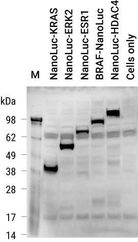
Detection of NanoLuc® fusion proteins expressed in HEK293 cells.
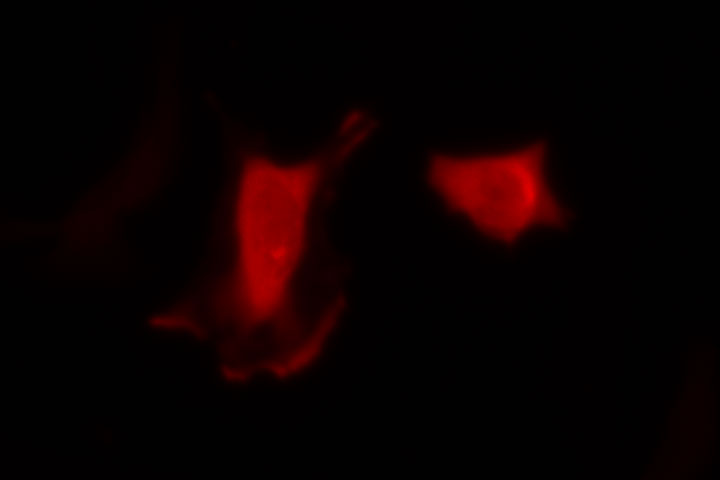
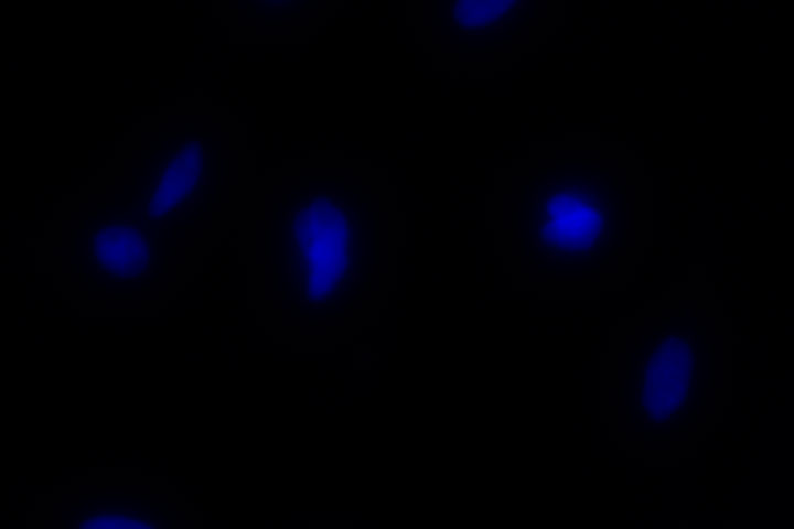
Immunofluorescence Results
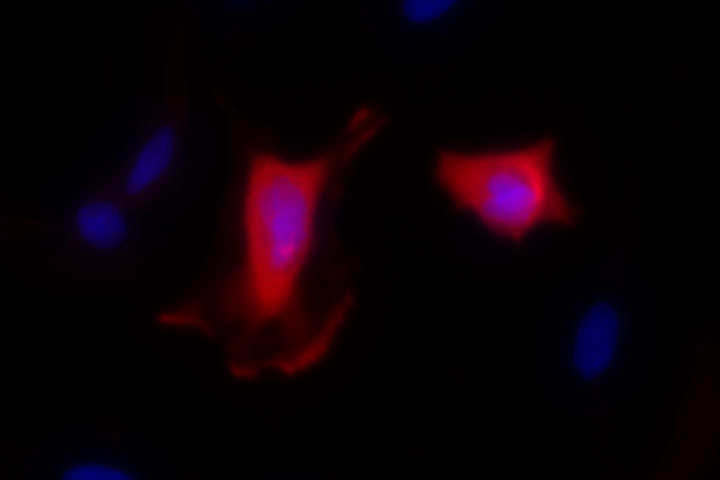
Shown below are example immunofluorescent images of HeLa cells transfected with a CMV-based construct expressing CDK2-NanoLuc® fusion protein. The CDK2-NanoLuc® fusion protein is stained red, and nuclei are marked blue via Hoechst staining. We recommend a concentration of 1-7.5µg/ml for immunofluorescence optimization. No NanoLuc® staining is observed in HeLa cells transfected with only carrier DNA (mock).
CDK2-NanoLuc®
(3µg/ml Anti-NanoLuc®)
NanoLuc®
Hoescht
Merge
Mock (5µg/ml Anti-NanoLuc®)
NanoLuc®

Hoescht
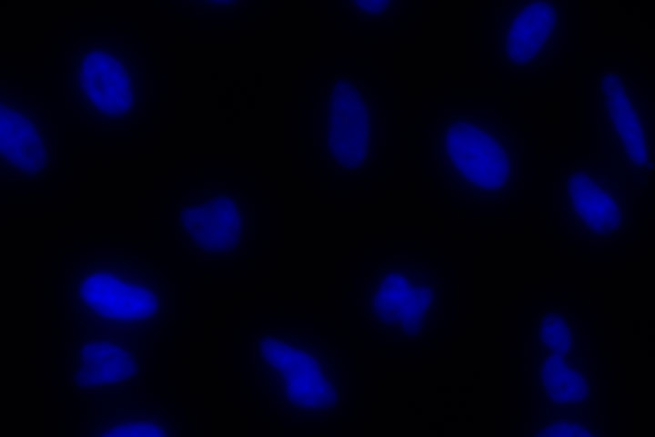
Merge
Protocols
Specifications
Catalog Number:
Contenido
| Item | Part # | Presentación |
|---|---|---|
Anti-NanoLuc® Monoclonal Antibody |
N700A | 1 × 100μg |
SDS
Search for SDSCertificado de Análisis
Use Restrictions
For Research Use Only. Not for Use in Diagnostic Procedures.Condiciones de Almacenaje
Related Products
Usado con frecuencia junto con
NanoBRET® PPI Starter Systems
Ensayo de interacción proteína:proteína en células vivas. Incluye vectores, controles y reactivos de detección para PPI basada en BRET.
N1811, N1821
NanoBRET® Target Engagement Kinase Assays
Permite medir de forma directa la afinidad de unión de la quinasa al ligando, la ocupación y el tiempo de residencia en células vivas.
N2520, N2521, N2540, N2501, N2500, N2530, N2600, N2601, N2620, N2621, N2630, N2631, N2640, N2641, N2650, N2651, NF1001, N2810, N2820, N2830, N2840, N2850, NF1200
Nano-Glo® Live Cell Assay
Mide la luminiscencia NanoBiT® o NanoLuc® en células vivas durante un máximo de 2 horas.
N2011, N2012, N2013
Nano-Glo® Luciferase Assay System
Ensayo "add-mix-measure" de NanoLuc® Luciferase con >2 horas de vida media en la mayoría de las aplicaciones.
N1110, N1120, N1130, N1150