LDH-Glo™ Cytotoxicity Assay
Simple, More Sensitive LDH Assay Method to Determine Cytotoxicity
- Detects LDH release from small numbers of cells, including 3D cell models
- Monitors cytotoxicity from the same sample over time
- Provides more data per well through multiplexing with other cell-based assays
- Integrates seamlessly with the MyGlo® Reagent Reader


Catalog Number:
Choose a product
Size
Catalog Number: J2380
Catalog Number: J2381
Monitor Cytotoxicity from Small Numbers of Cells
The LDH-Glo™ Cytotoxicity Assay is a bioluminescent plate-based assay for quantifying lactate dehydrogenase (LDH) release into the culture medium upon plasma membrane damage. The bioluminescent detection is more sensitive than colorimetric or fluorescent methods, allowing accurate detection of LDH from a small number of cells, including primary cells and 3D cell cultures.
This LDH assay involves removing only a small amount of cell media (2–5µl) from each treated well, allowing you to get more data by sampling the same well over time and by using the remaining media and cells for other cell-based assays.
How the Assay Works
LDH released from damaged cells catalyzes the oxidation of lactate with concomitant reduction of NAD+ to NADH. Reductase uses NADH and reductase substrate to generate luciferin, which is converted to a bioluminescent signal by Ultra-Glo™ Recombinant Luciferase. The luminescent signal generated is proportional to the amount of LDH present.
"The Promega LDH-Glo™ Assay is a reliable and easy-to-follow kit to use when measuring cell death. We use this in our lab, and compared to others, it really helps us get reliable data. Overall, we would highly recommend this kit."
LDH-Glo Customer
Monitor Cytotoxicity in 3D Cell Cultures
The LDH-Glo™ Cytotoxicity Assay is well-suited for measuring LDH released from small numbers of membrane-damaged cells. Here is an example of measuring drug-induced toxicity in HCT116 spheroids. The time-dependent toxicity measurements were performed by repeatedly sampling media from the same wells.
Detect Target Cell Cytotoxicity in ADCC Assays
The LDH-Glo™ Cytotoxicity assay can be used to assess cell death due to many mechanisms of action of biologics treatments, including complement-dependent cytotoxicity (CDC), antibody-dependent cell-mediated cytotoxicity (ADCC) and antibody-drug conjugate cytotoxicity. In this example, the LDH-Glo™ Assay was used to detect ADCC killing of target cells using the biologic rituximab.

Pair with the MyGlo® Reagent Reader
Reading your assay results doesn't require a shared plate reader. The MyGlo® Reagent Reader is a compact 96-well luminescence plate reader, enabling you to run luminescent assays at your bench without competing for instrument time. Integrated software generates dose response curves, single concentration and linearity analysis in minutes.

Protocols
Complete Protocol
Specifications
Catalog Number:
Contenido
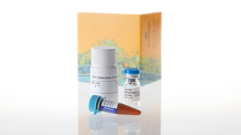
| Item | Part # | Presentación |
|---|---|---|
Reductase Substrate |
G885A | 1 × 55μl |
Lactate Dehydrogenase |
J195A | 1 × 1 each |
LDH Detection Enzyme Mix |
J238A | 1 × 10ml |
SDS
Search for SDSCertificado de Análisis
Use Restrictions
For Research Use Only. Not for Use in Diagnostic Procedures.Condiciones de Almacenaje
U.S. Pat. Nos. 9,273,343 and 9,951,372, European Pat. No. 2751089, Japanese Pat. No. 6067019 and other patents pending.
Contenido
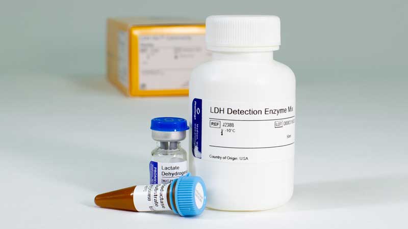
| Item | Part # | Presentación |
|---|---|---|
Reductase Substrate |
G885B | 1 × 275μl |
Lactate Dehydrogenase |
J195A | 1 × 1 each |
LDH Detection Enzyme Mix |
J238B | 1 × 50ml |
SDS
Search for SDSCertificado de Análisis
Use Restrictions
For Research Use Only. Not for Use in Diagnostic Procedures.Condiciones de Almacenaje
U.S. Pat. Nos. 9,273,343 and 9,951,372, European Pat. No. 2751089, Japanese Pat. No. 6067019 and other patents pending.
Resources
Artículos
- "Will This Drug Cause Hepatotoxicity?"
- App Note: Predicting gastrointestinal toxicity
- Impact of aerosols on liver xenobiotic metabolism: A comparison of two methods of exposure
- Folate and Vitamin B12 Deficiency Exacerbate Inflammation during Mycobacterium avium paratuberculosis (MAP) Infection.
- SARS‐CoV‐2‐reactive T‐cell receptors isolated from convalescent COVID‐19 patients confer potent T‐cell effector function
Related Products
Productos Similares
CellTox™ Green Cytotoxicity Assay
Mide los cambios en la integridad de la membrana. Monitoriza cinéticamente la citotoxicidad hasta 72 horas y además tiene capacidad multiplex.
G8741, G8742, G8743, G8731
CytoTox-Glo™ Cytotoxicity Assay
Ensayo de citotoxicidad basado en luminiscencia, de alta sensibilidad que mide el número relativo de células muertas.
G9290, G9291, G9292
CytoTox-ONE™ Homogeneous Membrane Integrity Assay
Método fluorométrico homogéneo basado en la LDH que le permite cuantificar las células no viables.
G7890, G7891, G7892
CytoTox-Fluor™ Cytotoxicity Assay
Ensayo fluorescente homogéneo de adición de un solo reactivo, que le permite medir el número relativo de células muertas en poblaciones celulares.
G9260, G9261, G9262
Usado con frecuencia junto con
CellTiter-Glo® 3D Cell Viability Assay
Método homogéneo optimizado para evaluar la viabilidad en cultivos celulares 3D.
G9681, G9682, G9683
GloMax® Discover System
Lector de microplacas de alto rendimiento para la detección de luminiscencia, fluorescencia y absorbancia.
GM3000
RealTime-Glo™ MT Cell Viability Assay
Método bioluminiscente que permite monitorizar la cinética de viabilidad de cultivos celulares durante 72 horas.
G9711, G9712, G9713
Caspase-Glo® 3/7 Assay System
Ensayo luminiscente fácil de usar que permite detectar la actividad de la caspasa-3/7.
G8090, G8091, G8093, G8092