Luminescence Based Assay for Proteasome Activity in Tissue Extracts
Publication Date: 2010
Abstract
Here we review a paper published in Analytical Biochemistry in which the Proteasome-Glo™ Assays were used to assay for proteasome activity in mouse skeletal muscle extracts.
Introduction
The ubiquitin-proteasome system (UPS) targets proteins for degradation, playing important roles in removing aberrantly synthesized or misfolded proteins and regulating protein half-life/activity in the cell. The UPS is implicated in regulation of the cell cycle(1), suppression of HPV oncogenesis in cervical cancer(2), and loss of proteasome function is implicated in some diseases including neurodegenerative diseases(3) , diabetes(4) and myofibrillar myopathies(5).
Because tissue samples from biopsy specimens are usually available in only small amounts, investigating proteasome-associated diseases is difficult. In a paper by Strucksberg et al., published in Analytical Biochemistry(6), the authors adapted a highly sensitive proteasome activity assay for use with small quantities of skeletal muscle. Bioluminescent proteasome assays have been developed for purified proteasome(7) (Proteasome-Glo™ Assays) and for cells in culture(8) (Proteasome-Glo™ Cell-Based Assays); this article describes an application of the Proteasome-Glo™ Assay for tissue extracts. Applying this sensitive and reliable assay to tissue extracts could enable more thorough investigation of the mechanism of proteasome-mediated pathologies.
"This article describes an application of the Proteasome-Glo™ Assay for tissue extracts. Applying this sensitive and reliable assay to tissue extracts could enable more thorough investigation of the mechanism of proteasome-mediated pathologies."
Tissue Preparation
The authors developed their assay using mouse quadriceps femoris and soleus muscles. In their work, they determined that tissue sample preparation is critical for reliable assay results. Tissues were sonicated and lysates prepared using ice cold reagents and cold centrifugation. Protein content was measured using a fluorescent quantitation kit. After quantitation, 3µg of the protein extract supernatants were analyzed via Western blot, and the amount of the 26S proteasome S4-subunit in each tissue extract was determined for normalizing activity assay results later.
Assay Protocol
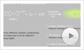
For the proteasome activity assay, the authors modified the Proteasome-Glo™ Chymotrypsin-Like Assay and the Proteasome-Glo™ Trypsin-Like Assay. These assays monitor two of the three catalytic activities of the proteasome, and were originally designed for applications such as screening compounds for their effects on proteasome activity rather than characterizing proteasome activity from tissue extracts. Then enzyme-coupled luminescent assay requires just a single addition step. The proteasome activity cleaves a luminogenic substrate containing a specific signal peptide coupled to aminoluciferin to yield luciferin. The released luciferin serves as a substrate for luciferase in a second, simultaneous reaction to generate light. This enzyme-coupled reaction produces a stable luminescent signal that is proportional to the proteosome activity in the sample.
To adapt the assays for tissue extracts the authors developed the following procedure:
- Extracts were prepared as described in the paper, and 3µg of extract supernatant was analyzed by Western blot to quantify the amount of 26S proteasome S4-subunit.
- Protein extracts were diluted to 0.2mg/ml in ice-cold PBSE.
- A total of 10µg of protein (50µl of diluted extract) was added to 50µl of the assay reagent containing the Ultra-Glo™ luciferase and the specific luminogenic substrate (Suc-LLVY-Glo™ for the chymotrypsin-like activity assay or Z-LRR-Glo™ for the trypsin-like activity assay) in a 96-well plate.
- Components were mixed and incubated for 60 minutes at room temperature.
- Luminescence signal was read three times with an integration time of 1 second using a Fluoroskan Ascent FL plate reader equipped with a count photomultiplier in the luminometry mode.
The luminescent signal intensity is a reflection of the total peptidase activity: that of the proteasome and any nonspecific peptidase present in the extract that might cleave the peptide from the aminoluciferin. To calculate proteasome activity, two measurements were made: one with the addition of the irreversible and specific proteasome inhibitor, adamantane-acetyl-(6-aminohexanoyl)3-(leucinyl)3-vinyl-(methyl)-sulfone, and one without inhibitor. Proteasome activity was calculated by subtracting the values obtained from the assays with inhibitor (background) from the total activity (no inhibitor). Proteasomal activity across different extracts was normalized to the amount of proteasome present in the extracts using the immunoblots performed earlier.
Considerations and Results
Several factors were critical for achieving a sensitive and reliable assay. The authors noted that even one cycle of freezing extracts at –80°C dramatically reduced proteasomal activity. Also, the amount of extract used affected signal intensity and stability and had to be optimized for this experimental system. Analysis of at least two mice was required to obtain reliable results with acceptable standard deviations.
The Proteasome-Glo™ Cell-Based Assays are designed to minimize the release of lysosomal proteases that can produce nonspecific background signal; however, with tissue extracts, lysosomal proteases are difficult to avoid particularly when sonicating samples. The authors use a proteasome-specific inhibitor to ensure specificity of the signal. Accounting for the nonspecific activity and normalizing to the total amount of proteasome in these extracts were essential steps for adapting these assays for use with tissue extracts. Once the assay was optimized for mouse skeletal muscle, the authors demonstrated that proteasome activity differs for different muscles within the same “normal” animal. They also showed that proteasome activity decreases with age.
Related Products
Related Protocols
 Learn about the chemistry of the bioluminescent reaction and its use in a broad range of assays.
Article References
- Cui, C. et al. (2010) Degradation of the human mitotic checkpoint kinase Mps1 is cell-cycle regulated by cdc20 and APC/cCdh1 ubiquitin ligases. J. Biol. Chem. 285, 32988–98.
- Alam, S. et al. (2006) Adeno-Associated Virus Type 2 increases proteasome-dependent degradation of p21WAF1 in a Human Papillomavirus Type31b-positive cervical carcinoma line. J. Virol. 80, 4927–39.
- Branco, et al. (2010) Crosstalk between mitochondria and proteasome in Parkinson disease pathogenesis. Frontiers in Aging, Neuroscience 2, 17.
- Hu, J. et al. (2010) Cardiac muscle protein catabolism in diabetes mellitus: Activation of the ubiquitin-proteasome system by insulin deficiency. Endocrinology 149, 5384–90.
- Schröder, R. and Schoser, B. (2009) Myofibrillar myopathies: A clinical and myopathological guide. Brain Pathol. 19, 483–92.
- Strucksberg, K.H. et al. (2010) Proteasomal activity in skeletal muscle: a matter of assay design, muscle type, and age. Anal. Biochem. 399, 225–9.
- O'Brien, M.A. et al. (2008) Homogeneous, bioluminescent proteasome assays. In: Apoptosis and Cancer: Methods and Protocols. Humana Press. Mor, G. and Alvero, A., eds. Methods in Molecular Biology Series 414, 163–81.
- Moravec, R.A. et al. (2009) Cell-based bioluminescent assays for all three proteasome activities in a homogeneous format. Anal. Biochem. 387, 294–302.
How to Cite This Article
Scientific Style and Format, 7th edition, 2006
Promega Corporation Luminescence Based Assay for Proteasome Activity in Tissue Extracts. [Internet] 2010. [cited: year, month, date]. Available from: https://www.promega.com/resources/pubhub/luminescence-based-assay-for-proteasome-activity-in-tissue-extracts/
American Medical Association, Manual of Style, 10th edition, 2007
Promega Corporation Luminescence Based Assay for Proteasome Activity in Tissue Extracts. Promega Corporation Web site. https://www.promega.com/resources/pubhub/luminescence-based-assay-for-proteasome-activity-in-tissue-extracts/ Updated 2010. Accessed Month Day, Year.
Proteasome-Glo, Suc-LLvY-Glo, Ultra-Glo and Z-LRR-Glo are trademarks of Promega Corporation.