Lumit® C1q Binding Assay
Evaluate C1q-antibody binding to assess the initiation of the complement-dependent cytotoxicity pathway
- Improved Immunoassay Format: ELISA alternative
- Saves Time: Reduce hands-on steps with a simple, no-wash protocol
- Better Data: No immobilization to plates, beads or other surfaces required
- Results in as little as 60 minutes
Catalog Number:
Size
Catalog Number: CS3695A04
The Ideal Assay to Complement Your Antibody Development Efforts
The Fc region of monoclonal antibodies mediate several effector functions, including complement-dependent cytotoxicity (CDC), through activation of the classical complement pathway. C1q binding to the Fc region is the critical first step in activating this pathway. By quantifying Fc-C1q binding, antibody developers can precisely profile C1q-antibody interactions, predict complement activation kinetics, and accelerate therapeutic antibody selection to ensure both efficacy and safety in therapeutic applications.
Traditional methods to measure C1q binding face several limitations:
- Time-consuming incubation steps
- Highly variable fluorescent readouts
- Low throughput assay set-ups
What does using the Lumit® C1q Binding Assay mean for you?
- Get simple, reliable surrogate measurement of CDC potential in under two hours
- Analyze multiple antibody isotypes and fine-tune your Fc engineering efforts and select promising candidates quickly
- Balance efficacy with safety: achieve sufficient complement activation while minimizing off-target toxicity
- Run your samples in a high-throughput and automation friendly design
How Does the Assay Work?

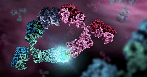
Principle of the Lumit® C1q Binding Assay: The assay provides a quantitative readout of C1q binding to antibody Fc as a proxy for classical complement engagement potential. In a homogeneous, solution-phase format, a biotinylated anti-human IgG Fab is pre-complexed with streptavidin–SmBiT to multivalently capture IgG and induce Fc clustering. C1q–LgBiT is then added; when C1q binds the clustered Fc domains, LgBiT and SmBiT are brought into proximity to reconstitute active NanoBiT® luciferase, producing a luminescent signal proportional to the amount of C1q-bound antibody.
What Is the Assay Workflow?

Lumit® C1q Binding Assay design:
- Biotinylated anti-human IgG Fab is pre-complexed with genetically fused Streptavidin-SmBiT, which multivalently captures IgG in solution and induces Fc clustering.
- Upon C1q-LgBiT addition and IgG binding, SmBiT and LgBiT are brought together, reconstituting active NanoBiT® luciferase.
- The luminescent signal is proportional to C1q bound to the antibody, enabling quantification of antibody-C1q interactions.
Accurately Detect Binding Differences Across Antibody Variants

Lumit® C1q binding assay is suited for distinguishing antibody specificity and sensitivity. The Lumit® C1q Binding Assay was tested against a panel of Rituximab variants that included different IgG isotypes as well as fucoslyation and glycosylation modifications. The binding profiles align with published literature.
Compare for yourself: Impact of structural modifications of IgG antibodies on effector functions.
| Rituximab | Non-fucosylated Rituximab | Non-Glycosylated Rituximab | Rituximab IgG2 isotype | Rituximab IgG3 isotype | Rituximab IgG4 isotype | |
| EC50 (nM) | 9.192 | 8.790 | 5278040 | 57.47 | 2.455 | 38534 |
Interested in other Fc effector functions like ADCC or ADCP?
We offer a wide range of Lumit® binding assays to measure interactions between Fc receptors and antibodies.Protocols
No protocols available
Specifications
Catalog Number:
Contenido
| Item | Part # | Presentación |
|---|---|---|
Human C1q-LgBiT |
CS3695A01 | 1 × 60μl |
Streptavidin-SmBiT |
CS3695A02 | 1 × 30μl |
Anti-human IgG Fab-Biotin |
CS3695A03 | 1 × 150μl |
Lumit® Immunoassay Dilution Buffer A, 10X |
VB201A | 1 × 10ml |
Lumit® Detection Substrate A |
VB301D | 1 × 75μl |
Resources
Featured Resource: Webinar
Lumit® Immunoassays: An Easier Faster Method for Analyte Detection
In Lumit® Immunoassays, antibodies (or other affinity reagents) are chemically labeled with the small and large subunits of NanoLuc® Luciferase, known as SmBiT and LgBiT, respectively. In the presence of an analyte, the two antibodies come into close proximity, allowing SmBiT and LgBiT to form an active enzyme and generate a bright luminescence signal.
- Use simple, add-mix-read protocols to detect a variety of analytes
- Implement existing Lumit® assays in your lab or build your own Lumit® immunoassays
- Analyze data using examples for cytokine detection, signaling pathway analysis and FcRn binding
Related Products
Productos Similares
Lumit® Immunoassay Cellular Systems
Inmunoensayo bioluminiscente sin lavado para medir los niveles de analitos diana en lisados celulares.
W1201, W1202, W1203, W1331, W1332, W1333, W1220
Lumit® Immunoassays para la detección de reguladores metabólicos
Ensayos para detectar insulina o glucagón en muestras de cultivos celulares con un protocolo escalable y sin lavado.
Usado con frecuencia junto con
Lumit® FcγR Binding Immunoassays
Nuevos ensayos homogéneos competitivos para medir la interacción entre receptores Fc humanos y anticuerpos o proteínas de fusión Fc.
W7030, W7031, W7040, W7041, W7050, W7051, W7060, W7061, W7070, W7071, W7080, W7081
Lumit® FcRn Binding Immunoassay
Un nuevo ensayo competitivo homogéneo (sin lavado) para medir la interacción entre el FcRn humano y las proteínas Fc, incluidos los anticuerpos.
W1151, W1152
ADCC Reporter Bioassay, V Variant
Measure potency and stability of antibodies mediating ADCC through the high-affinity human FcγRIIIa-V receptor.
G7015, G7010, G7016, G7018, G7102, GA1130
ADCP Reporter Bioassay (THP-1)
Measure the potency and stability of antibodies and other biologics with Fc domains that bind and activate FcγRs.
JA9411, JA9415, GA1272, GA6020