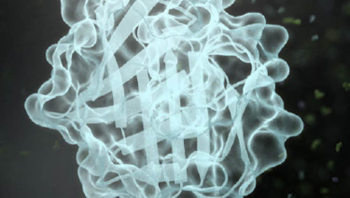
Bioluminescence Imaging
NanoLuc® Luciferase allows sensitive, bright bioluminescent imaging with low background and no excitation required.
Bioluminescent imaging of cells and molecular processes in whole animals provides important insights when studying normal physiology, monitoring disease progress or understanding response to therapy. NanoLuc® reporter technologies provide new tools for studying biological processes within whole animal models. These bright and small reporter options allow versatile in vivo applications ranging from quantifying changes in tumor growth to visualizing viral replication and spread using engineered NanoLuc® reporter viruses.
In Vivo Bioluminescence Imaging
NanoLuc® reporters provide high sensitivity and low background when imaged in superficial tissues and have also been used successfully to image events in deeper tissues. ATP-independence of these reporters allows in vivo monitoring of both intracellular and extracellular events.
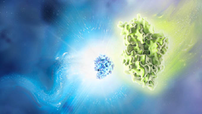
In addition, several in vivo imaging strategies have been developed using NanoLuc®-based BRET reporters. These techniques utilize the bright NanoLuc® signal to excite red-shifted fluorescent acceptor proteins, creating enhanced deep tissue imaging solutions.
The Nano-Glo® Fluorofurimazine In Vivo Substrate (FFz) is an optimized reagent designed specifically for in vivo detection of NanoLuc® Luciferase, NanoLuc® fusion proteins or reconstituted NanoBiT® Luciferase. This aqueous-soluble reagent provides increased substrate bioavailability in vivo, leading to bright signals, and has handling requirements compatible with in vivo workflows. In addition, substrate specificity allows NanoLuc® and firefly luciferases to be used together for dual-luciferase molecular imaging studies, providing even more options for creating whole animal reporter models.
In Vivo Bioluminescence Imaging Applications
Image the Brain of Living Animals with NanoLuc® Luciferase
Nano-Glo® Cephalofurimazine (CFz9) In Vivo Substrate enables the detection of NanoLuc® Luciferase in the brain of living animals. CFz9 was designed to cross the blood-brain barrier, enabling noninvasive exploration of central nervous system physiology and anatomy and studies of therapeutic distribution to the brain.
Learn more about the CFz9 substrate in this webinar.
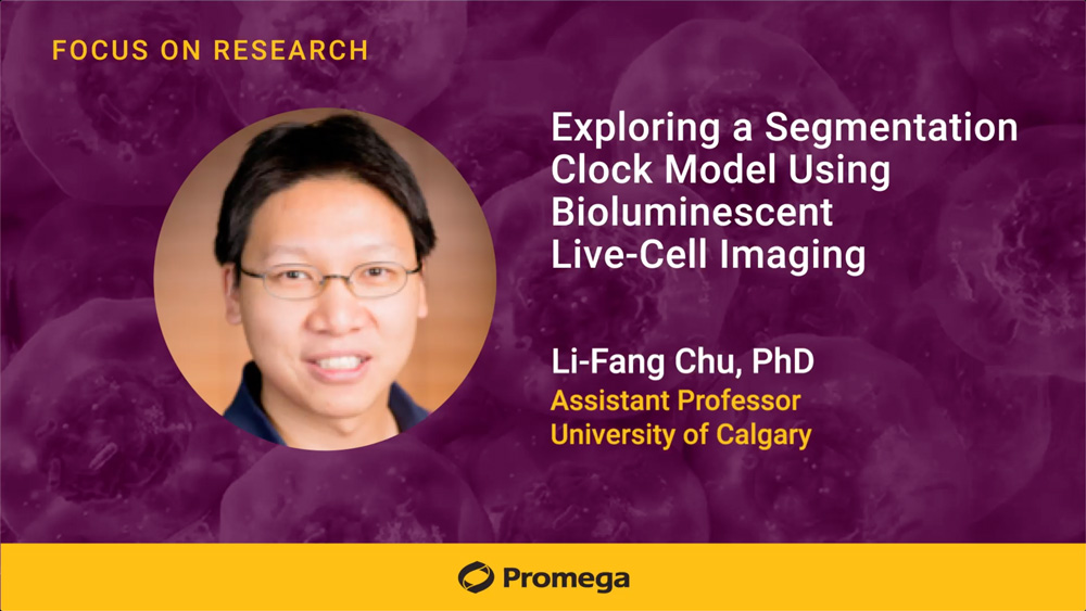
Pharmacodynamics of Akt drugs revealed by a kinase-modulated bioluminescent indicator
In Vivo Bioluminescence Imaging enables noninvasive monitoring of drug pharmacodynamics in real time. Bioluminescent indicators allow researchers to track drug activity, bioavailability, and duration of effect in living systems. This approach accelerates drug optimization for kinase inhibitors and other therapeutics while reducing preclinical testing requirements.
Bioluminescent Imaging in Live Cells
Control of subcellular localization is an important mechanism for regulating the function and signaling activity of many proteins. For example, protein translocation from the cytosol to the nucleus or protein recruitment to the plasma membrane can be key events in signaling pathway activation. Bioluminescent imaging (BLI) can be used to monitor subcellular protein localization, allowing direct visualization of protein dynamics in living cells without the need for repeated sample excitation.
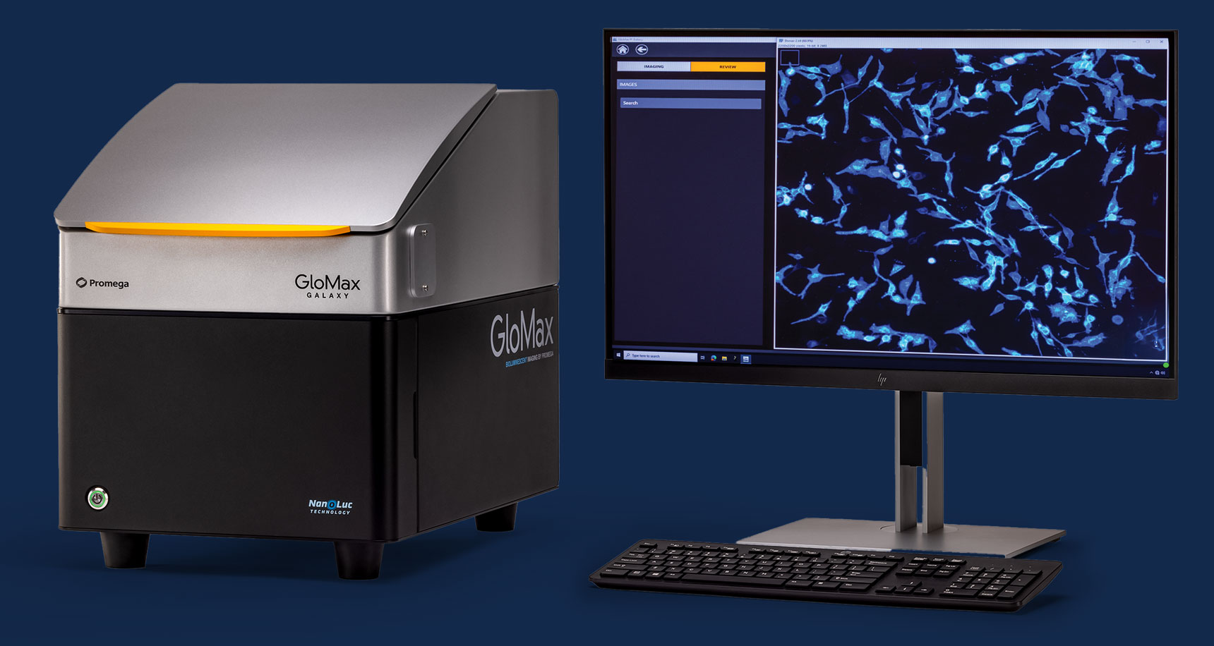
Live Cell Imaging System
To identify relevant biological processes, capturing images of live cells is a must. The GloMax® Galaxy Bioluminescence Imager combines the strength of bioluminescent reporters with the information acquired through live imaging. It is designed to enable imaging of NanoLuc® Luciferase technologies such as NanoBRET, NanoBiT, HiBiT and Lumit.

Live Cell Substrates
NanoLuc® Luciferase is well-suited for use as a protein tag in BLI studies. The extreme brightness means that exposure times can be reduced to only a few seconds, compared to the minutes required for other luminescent reporter proteins. In addition, its small size makes it less likely to perturb the normal biology or functionality of the fusion partner. Our Nano-Glo® Extended Live Cell Substrates have increased signal stability, allowing extended kinetic analysis lasting several hours to days.
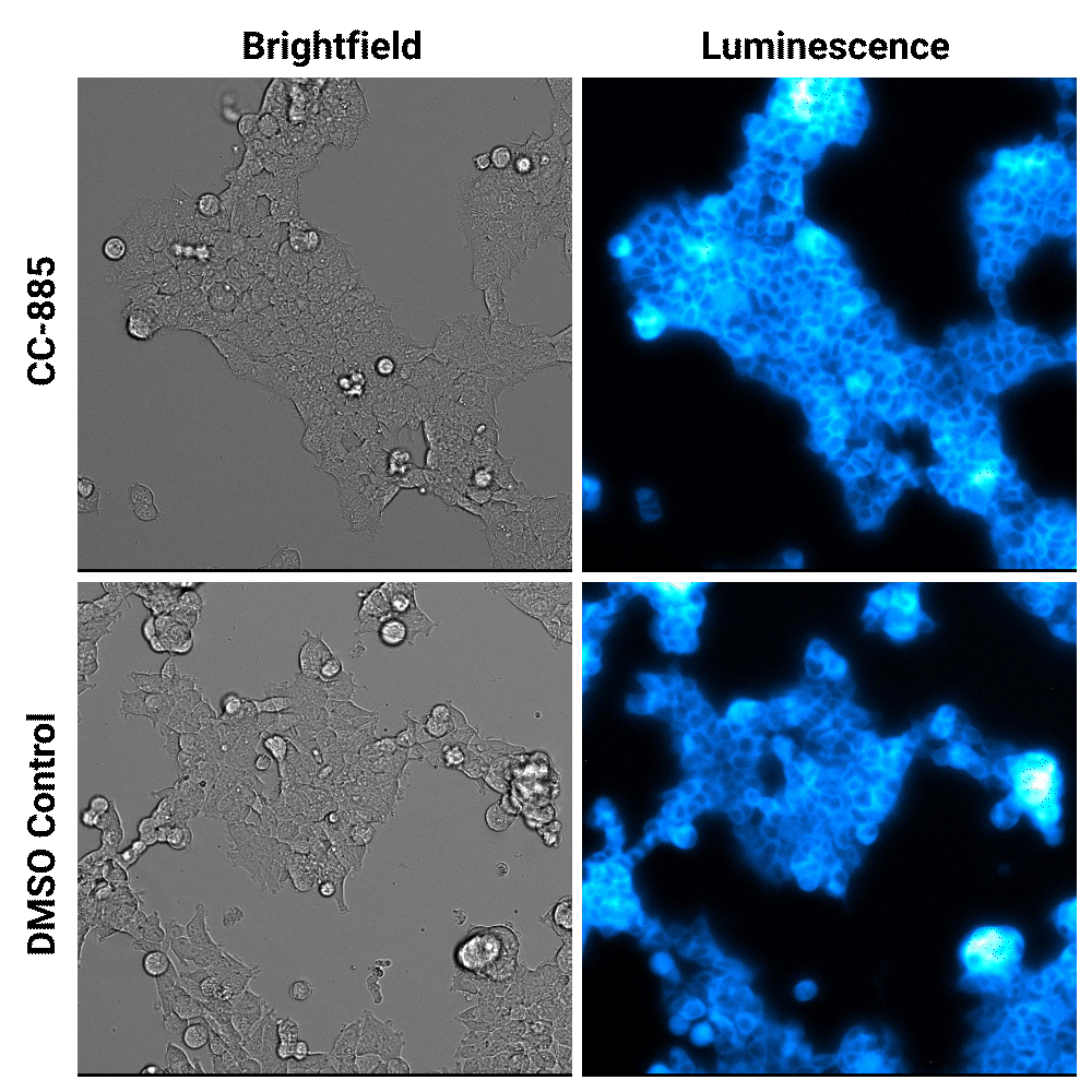
In the cell, things are very dynamic; they are happening every second. The bioluminescence live-cell imaging was what allowed us to discover this dynamic"
Researchers in Dr. Chu's lab observed the exact timing of oscillating genes in early human development for the first time using bioluminescence live-cell imaging. You can read more about this work in the blog: Observing the human developmental clock with bioluminescence live-cell imaging.

Featured Lab Manager Article: Unlocking Cellular Insights with a Bioluminescence Imaging System
This article outlines how bioluminescence imaging systems enhance experimental insight and highlights the critical features to evaluate when choosing the right platform for your research.
Explore Bioluminescent Imaging Applications
See how researchers are using the GloMax® Galaxy Bioluminescence Imager to get more from their NanoLuc® Luciferase experiments. Explore proven imaging applications and find inspiration for your next discovery.
Additional Resources
NanoLuc® Luciferase: Brighter Days Ahead for In Vivo Imaging
Read more about in vivo applications of NanoLuc® technology in this blog.
A Small Luciferase Brightening Up the Field of Bioluminescence
This review article details the advantages of NanoLuc® technology for the scientific community.
Novel Substrates Enable Two-Population In Vivo Bioluminescence Imaging
Read about the development of NanoLuc® substrates in this publication.
Learn More About Imaging
Imaging
Learn about various imaging techniques, including live cell imaging, in vivo imaging, bioluminescence imaging and fluorescence microscopy.
In Vivo Imaging
Learn about in vivo imaging techniques including bioluminescence and fluorescence imaging, advantages, challenges, and applications in research.
Live Cell Imaging
Learn about live cell imaging techniques, advantages and applications.