Beyond Viability: Functional Profiling in Perifusion-Based Islet Studies
Simon Moe, PhD. and Aisosa Omere
Publication Date: September 2026
Why Viability May Not Give You a Complete Picture
Rates of obesity have increased to unprecedented levels, with some studies classifying over 60% of populations as obese (Fourman, 2025). Classified as higher rates of Body Mass Index (BMI), obesity has been associated with elevated rates of diabetes. Teasing apart the underlying biology resulting in and from these disease states has continued to grow as these rates have increased over the past few decades.
A common tool for mapping metabolic dysfunction is perifusion models, where medium is continuously flowing over islet cells. Media is collected after it exits the chamber, providing a consistent sample to measure insulin, glucagon, and metabolites. Within diabetes and obesity research, cells can be alive yet metabolically incompetent. Insulin resistance, impaired glucose handling, and mitochondrial dysfunction are established features of type 2 diabetes (Prasun, 2020; Sergi et al., 2019), yet none of these characteristics show up in a viability screen. No assay is fully comprehensive, and as such, both viability and functional assays belong in a metabolic screen.
Perifusion captures islet secretion dynamics in real time, a valuable tool to understand how metabolic diseases impact secretion of factors such as insulin or glucagon. Within these perifusion models, screening for viability is crucial to understand dead cells aren't skewing your functional readouts. Viability functions as a contextual gate, within the context of a larger functional workflow.
How Viability and Functional Measures Work Together in Perifusion
How Do I Measure Viability in a Perifusion Context?
Perifusion collects supernatant fractions continuously, but you need to know how many live cells are secreting those fractions. The gold-standard endpoint assay for ATP-based luminescence detection is CellTiter-Glo® 2.0 Cell Viability Assay. CellTiter-Glo® 2.0 lyses the cells, and a luciferase reaction turns the released ATP into light. If you want a non-endpoint assay, the RealTime-Glo™ MT Cell Viability Assay enables same-well measurement of viability while the perifusion is occurring.
One caveat matters in metabolic work: ATP per cell is not fixed. Take a compound that impairs oxidative phosphorylation and it will lower the signal from CellTiter-Glo without killing anything. Before calling your experiment at a loss of CellTiter-Glo signal, consider corroborating it with a non-metabolic death measure, like CellTox™ Green Cytotoxicity Assay or LDH-Glo™ Cytotoxicity Assay, as is standard within some industrial cascades (Mervin et al., 2016).
How Do I Measure Metabolism and Hormone Secretion in a Perifusion Context?
As medium flows over the islets, you collect supernatant fractions at various timed intervals. These fractions contain various secreted factors. Due to the sensitivity of Promega metabolic activity assays, analysis can be performed on cell culture supernatants without having to damage the cells, allowing you to measure glucose consumption with the Glucose-Glo™ Assay and lactate secretion with the Lactate-Glo™ Assay. To measure insulin or glucagon secretion from your islets, The Lumit® Insulin Immunoassay and Lumit® Glucagon Immunoassay enable sensitive, no-wash bioluminescent detection.
Viability and Functional Assays for Perifusion Studies
| Assay | Readout | Lytic? | When to Use | Compatible with Perifusion Fractions? |
|---|---|---|---|---|
| CellTiter-Glo® 2.0 Cell Viability Assay | ATP-based cell viability | Yes | Endpoint viability | No, run on cells not fractions |
| RealTime-Glo™ MT Cell Viability Assay | Reducing potential (viability proxy) | No | Baseline viability before perifusion | No, run on cells not fractions |
| CellTox™ Green Cytotoxicity Assay | Membrane integrity/cell death | No | Corroborate unexpected CellTiter-Glo signal drops | No, run on cells not fractions |
| LDH-Glo™ Cytotoxicity Assay | LDH release/cytotoxicity | No | Confirm cell death independent of metabolic status | Yes, from collected fractions |
| Glucose-Glo™ Assay | Glucose concentration | No | Confirm stimulus, track substrate availability | Yes, from collected fractions |
| Lactate-Glo™ Assay | Lactate secretion | No | Glycolytic output during perifusion | Yes, from collected fractions |
| Lumit® Insulin Immunoassay | Insulin secretion | No | Beta-cell secretion kinetics | Yes, from collected fractions |
| Lumit® Glucagon Immunoassay | Glucagon secretion | No | Alpha-cell secretion kinetics | Yes, from collected fractions |
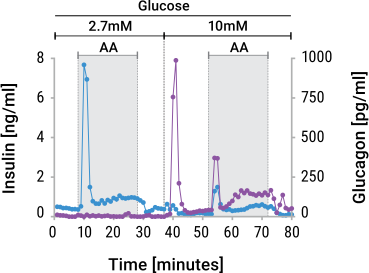
Measuring these secreted factors in a perifusion context provides you with time-resolved data from your sample. You can identify which secretion phase correlates with robust glucose utilization, or even where a metabolic state lags or leads to a certain hormone output. Contextualize these findings with viability to normalize for cell count. This ensures that when you see a drop in insulin signal, it means impaired secretion per cell, not fewer live cells.
How Do I Measure Viability and Metabolic Activity in Perifusion Experiments?
Example Workflow 1: Establish viability ceiling, then perifuse at matched doses
Best for: dose-response studies where compound toxicity is unknown
The first way to set-up this experiment would be to run a dose-response of your test compound on islets. You can measure viability using CellTiter-Glo 2.0 at each dose to find the highest concentration where cells stay >80% viable. This ceiling becomes your working range for functional studies. Now you can perifuse islets at those ceiling-matched doses and know that viability should remain stable. Collect supernatant fractions and measure Lumit® Insulin and Lumit® Glucagon kinetics alongside any metabolites on the same fractions, such as glucose consumption and lactate.
Example Workflow 2: Non-lytic viability baseline, then perifuse with fraction sampling
Best for: studies where preserving your islet preparation before perifusion is a priority
Before perifusion, measure viability non-lytically using RealTime-Glo® MT on your baseline islet preparation. This establishes cell count without destroying the cells you're about to perifuse. As medium flows over the islets during perifusion, collect supernatant fractions at various times. From those fractions, measure Lumit® Insulin/Glucagon and metabolites from aliquots of the same sample. If viability remains stable across your dose range, changes in Lumit® Insulin/Glucagon or metabolite output reflect functional impairment, not cell loss. The non-lytic read ensures that your experimental population is intact.
Why Should I Build a Metabolic Activity Screen Workflow?
Cost and Dose Selection
Utilizing cell viability or cytotoxicity as an initial gate pushes forward only the most likely candidates for the more expensive functional tier. It tells you which compound doses are safe enough to carry forward to perifusion, which is more labor-intensive and reagent-expensive than a static viability plate. This point is well highlighted when Mervin et al used a screen to narrow 388,000 compounds down to 5784 active cytotoxic hits in a workflow that utilized the CellTox™ Green Cytotoxicity Assay (Mervin et al., 2016). It would be impractical to conduct a more comprehensive screen on over 388,000 compounds to any notable degree. If viability is run concurrently during your perifusion experiments, it enables you to normalize your findings and be more confident on the dose selection.
Interpretability of Kinetics
Perifusion captures the complex kinetics of insulin activity, including a first-phase insulin spike, a second-phase plateau, and glucagon suppression. A raw, non-normalized value can be ambiguous: a 50% drop could mean impaired insulin secretion, or simply half as many cells still alive. Normalizing boosts confidence that the kinetic changes you see aren’t due to a drop in viability.
Mechanistic Distinction
Viability data lets you separate true beta-cell dysfunction from islet degeneration in obesity models. A perifusion experiment that has blunted first-phase insulin, but stable lactate output suggests secretion-specific impairment, likely either a mitochondrial or signaling defect. The same blunted insulin with significantly reduced lactate and glucose consumption suggests metabolic collapse. Without a viability confirmation throughout, you cannot rule out that cells simply died and stopped performing. Viability helps normalize your interpretation of which pathway broke in the islet.
In Summary
Viability is not the endpoint of a metabolic screen. It is the entry criterion and internal control. Run it first, on its own plate, to set a per-compound ceiling; then again in-well, ahead of a terminal functional read, to make the result per-cell. A perifusion experiment enables vital functional data to be collected, and viability strengthens those findings. Used this way, it becomes the thing that makes the functional result interpretable.
References
- Prasun, P. et al. (2020) Role of mitochondria in pathogenesis of type 2 diabetes mellitus. J. Diabetes Metab. Disord. 19, 2017–22.
- Sergi, D. et al. (2019) Mitochondrial (dys)function and insulin resistance: from pathophysiological molecular mechanisms to the impact of diet. Front. Physiol. 10, 532.
- Mervin, L.H. et al. (2016) Understanding cytotoxicity and cytostaticity in a high-throughput screening collection. ACS Chem. Biol. 11, 3007–23.


Learn more about our Cell Health Assays for measuring viability, cytotoxicity and metabolic activity