In Vivo Imaging
Learn about in vivo imaging techniques including bioluminescence and fluorescence imaging, advantages, challenges, and applications in research.
What Is In Vivo Imaging?
In vivo imaging refers to a variety of techniques used to visualize and study biological processes in whole, living organisms. It can be used to observe anatomical structures, molecular/cellular activities, and metabolic functions as they occur more naturally within whole organisms. This is different from in vitro imaging, which refers to techniques used to examine processes in cultured cells.
Advantages of in vivo imaging:
- Results taken from live animals are more biologically relevant
- Real-time monitoring of animals can provide insights into dynamic events, such as tumor development
Challenges of in vivo imaging:
- Higher cost of imaging equipment and animal care
- Requires larger sample sizes and longer timelines
Featured Products
HaloTag® Ligands for Imaging
Brighter, more photostable ligands for standard confocal imaging and high-resolution imaging in live cells.
Nano-Glo® Fluorofurimazine In Vivo Substrate (FFz)
Aqueous-soluble in vivo detection reagent with increased substrate bioavailability for bright, stable signals.
VivoGlo™ Luciferin
Firefly luciferase substrate for use in animal imaging studies. Tested to ensure low endotoxin levels for optimum performance in vivo.
In Vivo Bioluminescence Imaging
In vivo bioluminescence imaging (BLI) is a non-invasive technique used for real-time monitoring of cellular and molecular processes within live organisms. Unlike other imaging modalities that may require the use of contrast agents or radiation, BLI relies solely on the light produced by bioluminescence reaction.
Advantages of In Vivo Bioluminescence Imaging
- Non-toxic: Subjects can be repeatedly imaged over time without significant harm. This allows for longitudinal studies on disease progression and gene expression patterns.
- High sensitivity: The luciferase-luciferin reaction is highly efficient, producing a strong light signal with minimal background noise. This sensitivity enables the detection of very low levels of biological activity.
How Does In Vivo Bioluminescence Imaging Work?
In vivo BLI requires three main components:
- Animal model expressing a luciferase reporter gene
- Luciferase substrate
- Imaging system
The general process of in vivo BLI is as follows:
- Reporter animal model creation: An animal model expressing luciferase reporters, such as NanoLuc® or firefly luciferase, is created using genetic engineering techniques.
- Substrate administration: Luciferase substrate is administered into the animal, typically via injection.
- Luminescence signal generation: When the substrate encounters luciferase in the presence of oxygen and ATP, it undergoes an enzymatic reaction that produces luminescence.
- Detection and imaging: Specialized imaging systems detect the luminescence signal and convert it into an image.
Reporters and Substrates for In Vivo Bioluminescence Imaging
The luminescence signal generated from luciferase reporters is dependent on the substrate, so selecting an appropriate substrate is crucial. We provide substrates optimized for the following reporters:
- NanoLuc® Luciferase Detection: The Nano-Glo® Fluorofurimazine In Vivo Substrate (FFz) is designed specifically for in vivo detection of NanoLuc® Luciferase, NanoLuc® fusion proteins or reconstituted NanoBiT® Luciferase.
- Firefly Luciferase Detection: VivoGlo™ Luciferin, In Vivo Grade is optimized for in vivo detection of firefly luciferase, firefly luciferase fusion proteins, or reconstituted firefly luciferase complexes.
- NanoLuc® and Firefly Luciferase Dual Detection: Specificity of the above substrates allows NanoLuc® and firefly luciferases to be used together for dual-luciferase molecular imaging studies, providing even more options for creating whole animal reporter models.
Interested in our substrates designed for in vivo imaging?
Image the Brain of Living Animals with NanoLuc® Luciferase
Nano-Glo® Cephalofurimazine (CFz9) In Vivo Substrate enables the detection of NanoLuc® Luciferase in the brain of living animals. CFz9 was designed to cross the blood-brain barrier, enabling noninvasive exploration of central nervous system physiology and anatomy and studies of therapeutic distribution to the brain.
Learn more about the CFz9 substrate in this webinar.
In Vivo Bioluminescence Imaging Applications
Study Infectious Disease Dynamics
By using bioluminescent bacteria or viruses, researchers can non-invasively monitor the spread and localization of an infection in real-time within a living organism. This allows for the assessment of pathogen load, dissemination, and the host’s immune response over time, providing insights into disease mechanisms and the effectiveness of therapeutic interventions.
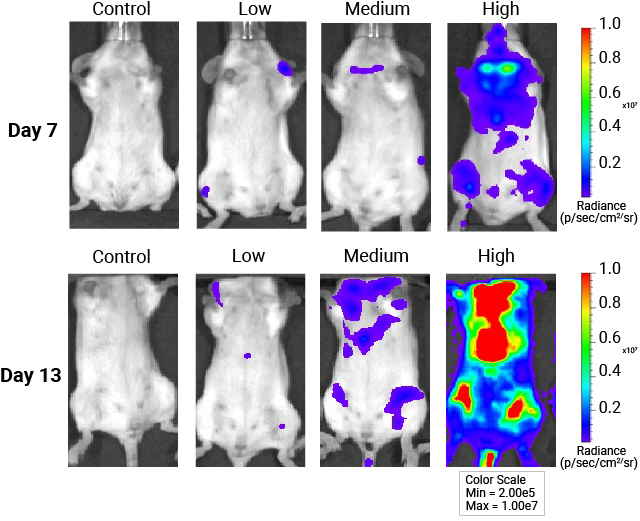
Cancer Progression
Tumor cells engineered to express luciferase can be tracked in vivo, allowing researchers to visualize and quantify tumor burden and metastatic spread longitudinally. This technique can be used to evaluate the efficacy of anti-cancer therapies, monitor tumor relapse, and study the biology of cancer cells in their native microenvironment.
Evaluate Gene Therapy Outcomes
By incorporating luciferase reporters into gene therapy vectors, researchers can assess the efficiency of gene delivery, expression levels, and the persistence of the therapeutic gene non-invasively over time. Real-time monitoring helps optimize gene therapy strategies, ensuring targeted and sustained gene expression.
Develop New Drugs and Therapies
In vivo BLI enables real-time evaluation of drug efficacy, pharmacokinetics, and biodistribution in live animals. This enables researchers to rapidly screen and identify promising therapeutic candidates, study drug mechanisms, and optimize dosing regimens.
In Vivo Fluorescence Imaging
In vivo fluorescence imaging (FLI) utilizes fluorescent probes or ligands, which emit light upon excitation by an external light source.
Advantages of In Vivo Fluorescence Imaging
- Multiplexing: Fluorescent probes are widely available for identification of protein and gene targets, enabling a wide variety of multiplexing possibilities.
- Quick Analysis: Due to the high number of photons produced by fluorescent ligands, images can be captured with short exposure times, enabling quick analysis of fluorescent markers.
Disadvantages of In Vivo Fluorescence Imaging
- Autofluorescence: Natural fluorescence from tissues can interfere with the signal from fluorescent probes, reducing contrast and accuracy.
- Photobleaching: Prolonged exposure to light can cause fluorescent molecules to lose their ability to emit light, diminishing the quality of imaging over time. Thus, long-term kinetic studies may not be possible.
- Toxicity: Some fluorescent dyes or probes can be toxic to living tissues, affecting the viability of the sample and the accuracy of the results.
In Vivo HaloTag® Ligands for Imaging
Our Janelia Fluor® HaloTag® Ligands, including JFX dyes, are engineered for characterization of HaloTag® fusion proteins in live-cell and endogenous environments. These dyes span the visible spectrum and exhibit unique fluorogenic properties, enhancing brightness and reducing background signal for high-resolution imaging applications.
Brain-wide measurement of synaptic protein turnover reveals localized plasticity during learning | bioRxiv
Product Citations
Resources
La Importancia de MSI
Aprenda sobre los usos de MSI como biomarcador de referencia en oncología y como apoyo en la toma de decisiones sobre el tratamiento.
Métodos de detección
Compare los diferentes métodos empleados para analizar MSI y/o MMR.
Poster: Assessment of AAV Biodistribution in Mice
Learn how the NanoLuc® Reporter can provide a simple, single day workflow that enables high-throughput viral titer determination of AAV infected tissues in live animals.