"Will This Drug Cause Hepatotoxicity?"
Getting answers by combining 3D cell culture with the right assay
October 01, 2018
Monika Kijanska, Ph.D, is on a mission to develop and provide 3D culture solutions for testing the safety and efficacy of drugs. She is a senior scientist and team lead of Liver Solutions at InSphero, a biotech company specializing in 3D cell culture technology for drug safety and efficacy testing.
Traditionally, drug safety testing was often done using 2D cultures grown flat in a dish, even though they don’t accurately represent living organs. More recently, 3D cultures (also known as spheroids and microtissues) have become the in vitro model of choice for drug screening and development. With 3D microtissue technology, you can co-culture different cell types and add organ-specific cell components to better mimic the cellular composition of a living organ. As a result, the functionality of 3D cultures can be preserved for much longer than in 2D models. This long lifespan is important for determining potential drug-induced liver injury (DILI). Certain compounds might have chronic effects in the liver that only occur after long exposure times, which might be missed in short-term 2D cultures that are metabolically competent only for a few days.
Although 3D cultures have many advantages, developing reliable and uniform 3D models for drug screening purposes is a slow and costly task for pharmaceutical companies. This is where Dr. Kijanska’s and the liver team at InSphero can provide their service and expertise.

Dr. Monika Kijanska
“Hepatoxicity is one of the major questions that we are trying to answer.”
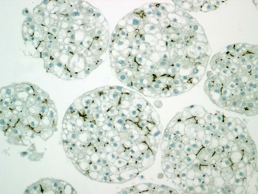
The InSphero liver team has developed 3D InSight™ Human Liver Microtissue models, which are highly standardized and uniform liver spheroids, cultured in specially designed 96-well plates. The key feature of these liver models is their enhanced metabolic competence and stability over up to 28 days in culture. To determine whether specific compounds cause long-term hepatotoxicity, these liver microtissues are treated with the compounds for 14 days and re-dosed multiple times during that period. At the end of that exposure, the overall viability is determined using an endpoint cell viability assay.
To evaluate whether drugs will cause DILI, researchers not only need to know the number of viable cells at the end of treatment, they also want to understand the kinetics of the cytotoxic effect. At what specific time and concentration did toxicity occur? The only way to get those data using traditional endpoint cytotoxicity assays is to set up separate plates of liver microtissues at each time point. This drastically increases the cost and time required to get answers.
“In order to solve most cell-based scientific questions, you need both a model and an assay.”
InSphero’s team set out to find the best cytotoxicity assay that could deliver accurate endpoint data for 3D InSight™ Liver Microtissues. Their ideal cytotoxicity assay would allow them to monitor cell death over a 14-day time course. It would allow them to multiplex with other assays to get as much data as possible in a cost-effective manner. It would be sensitive enough to detect cell death from a single microtissue. It would be scalable for use in both 96-well or 384-well formats. And finally, it would be fast and simple to run.
“Having a partner that provides assays to match our models is what we need to get the right answer.”
Through communication with Promega scientists Dr. Jolanta Vidugiriene and Dr. Natasha Karassina, InSphero’s liver team became one of the first to test a new cytotoxicity assay based on LDH release. Their team had already been using the well-established Promega CellTiter-Glo® Assay, which detects ATP as a marker for measuring cell viability, and they loved how easy it was to use. The protocol involved adding a reagent into sample wells containing cells, a short 10-minute incubation and direct measurement using a plate-reading luminometer. The simple “add-mix-measure” procedure is typical of Promega cell-based assays, and InSphero’s team wanted something similar that they could use for measuring cytotoxicity in their 3D models.
“LDH release has a good correlation with the cytotoxic compounds that we have tested so far. It really helps answer that question about the potential cytotoxicity of compounds.”
Lactate dehydrogenase (LDH) is a stable enzyme that rapidly leaks from the cell once the plasma membrane is disrupted, which happens upon cell death. For years, LDH has been widely used as a marker for cell death—but there was one common problem. Most existing assays use colorimetric or fluorometric methods for detection, which are often not sensitive enough to detect LDH released from single spheroids.
“We need only a few microliters of supernatant for the LDH-Glo™ Assay. That’s very sensitive.”
The new LDH-Glo™ Cytotoxicity Assay uses highly sensitive bioluminescence to quantify LDH release, allowing accurate detection of LDH release from a small number of cells. In fact, the assay is so sensitive that only a small amount of culture medium is needed for detection. This is especially valuable when working with individual 3D microtissues that are made of only a few thousand cells.
“It’s an easy assay. You just need to mix the supernatant with the reagent, then incubation and readout. It’s very straightforward.”
The LDH-Glo™ Assay involves removing 2–5µl of culture medium from the sample well, then diluting it in storage buffer, which preserves LDH activity. This gives you the flexibility to measure LDH release immediately or store the sample at –20°C for future detection without loss of LDH activity. When ready for detection, simply add the LDH detection reagent to the sampled medium and measure luminescent signal using a plate-reading luminometer. That’s it.
“We can get way more data from a single experiment with this LDH assay.”
Because only a few microliters of medium are needed for LDH detection, the medium can be repeatedly sampled from the same well throughout a time course. Samples can also be taken during medium changes. This eliminates the need to set up multiple plates for each time point. Today, Dr. Kijanska uses the LDH-Glo™ Assay to determine at what time and concentration a cytotoxic reaction takes place throughout the 14-day multi-dosing period. At the end of the exposure, they can still use the remaining cells and medium for other endpoint assays, such as cell viability or cytokine detection.
“The best assay is the one that helps you answer your research question.”
By pairing cell viability assays with the LDH-Glo™ Assay, Dr. Kijanska can get more definitive answers than an ATP-based cell viability assay can provide alone. For example, when ATP levels go down, it could be due to either metabolic stress or membrane breakage. Only when paired with an LDH assay can you get a definitive answer: Low ATP levels with high LDH levels indicate membrane breakage and necrosis; low ATP levels with low LDH levels indicate metabolic stress.
“We use the LDH-Glo™ Assay more and more on a routine basis for toxicity testing in liver microtissues. I also think it will work well with our other microtissue models.”
The LDH-Glo™ Assay works exceptionally well in 3D InSight™ Liver Microtissues, partly because InSphero’s specialized culture medium does not contain serum, so the background LDH level is very low. This may not be the case for other 3D spheroid models. Nonetheless, Dr. Kijanska is spreading the word about the LDH-Glo™ Assay to other teams at InSphero, and they expect that the assay will work well in InSphero’s other 3D models. She believes that their 3D model, combined with the LDH-Glo™ Assay, are an ideal combination for evaluating hepatotoxic effects of drugs.
Learn more about the LDH-Glo™ Cytotoxicity Assay.
Want to see the data?
Learn how the LDH-Glo™ Cytotoxicity Assay is used with different model systems, including SKBR3 cells, to assess an antibody drug conjugate and in 3D human liver microtissues to monitor cytotoxicity over time.